Sensory Cell Biology and Organogenesis
Explore CRG Images
The aim of the CRG photo gallery is to provide an overview of our scientists’ research activities through the images generated in the course of their research projects.
All our images are available on demand in digital form with higher quality for for-profit and any other uses. For any of these requests please contact us.
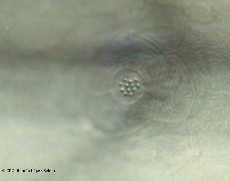
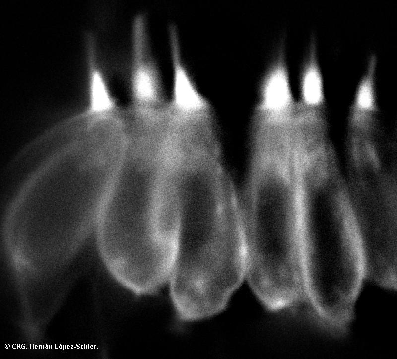
Title: Lateral line from zebrafish
Ref: HLS-0028
Author: HERNÁN LÓPEZ-SCHIER
Size: 800 x 630
Description: DIC (Differential interference contrast) front image of a lateral line organ of the zebrafish
Explore CRG Images
The aim of the CRG photo gallery is to provide an overview of our scientists’ research activities through the images generated in the course of their research projects.
All our images are available on demand in digital form with higher quality for for-profit and any other uses. For any of these requests please contact us.
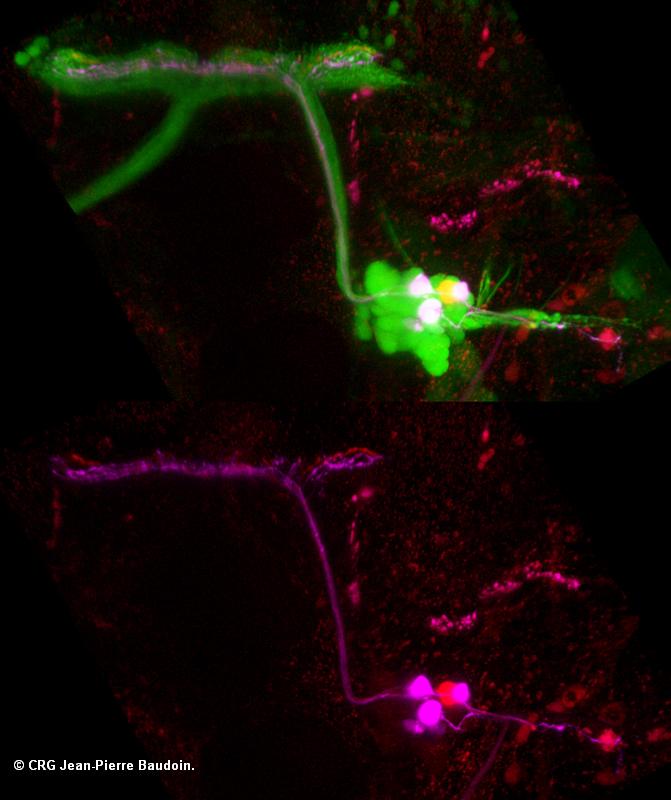
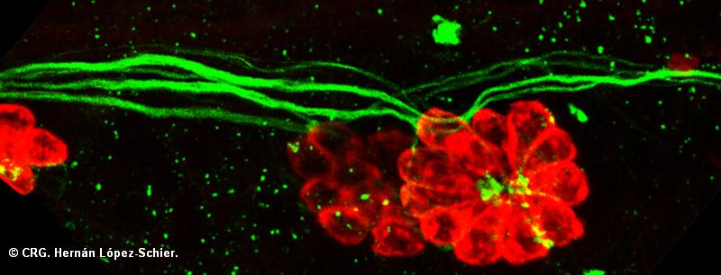
Title: Afferent neurons from zebrafish lateral line
Ref: HLS-0027
Author: JEAN-PIERRE BAUDOIN
Size: 671 x 800
Description: Scatter-label of afferent neurons of the lateral line by double injection of rhodamin- and magenta-dextran in a neuromast (red/magenta) of a zebrafish transgenic line HGn39D from K. Kawakami) (green)
Explore CRG Images
The aim of the CRG photo gallery is to provide an overview of our scientists’ research activities through the images generated in the course of their research projects.
All our images are available on demand in digital form with higher quality for for-profit and any other uses. For any of these requests please contact us.
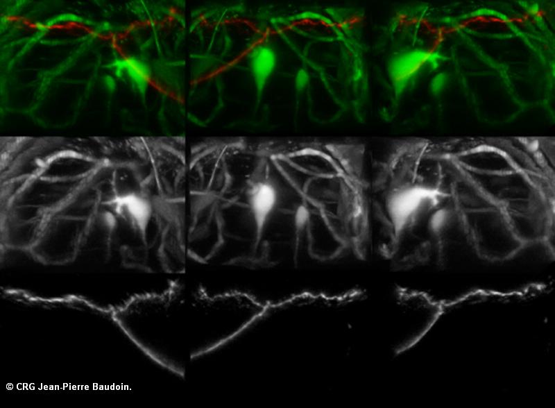
Title: 3D perspective of Mauthner cells from zebrafish
Ref: HLS-0026
Author: JEAN-PIERRE BAUDOIN
Size: 800 x 588
Description: Three angle perspective of a 3D rendering of a Mauthner cells (zebrafish transgeic line DMC130A from K. Kawakmi) (green) with a single neurons of the lateral line labelled by injcetion of rhodamin-dextran in a neuromast (red)
Explore CRG Images
The aim of the CRG photo gallery is to provide an overview of our scientists’ research activities through the images generated in the course of their research projects.
All our images are available on demand in digital form with higher quality for for-profit and any other uses. For any of these requests please contact us.
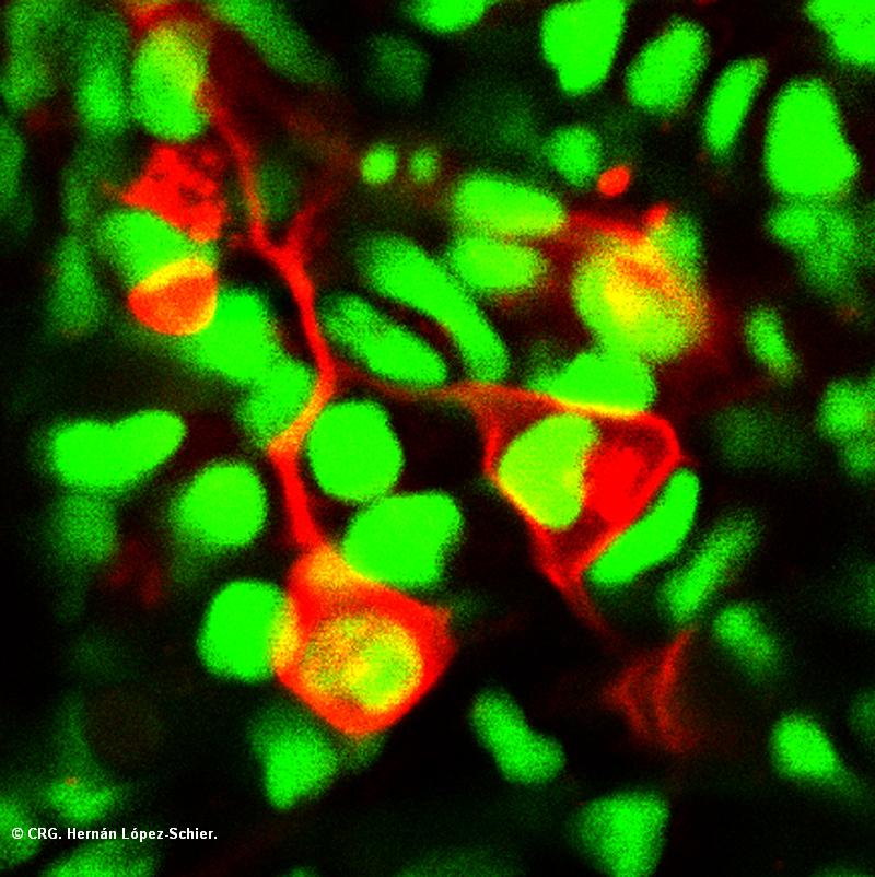
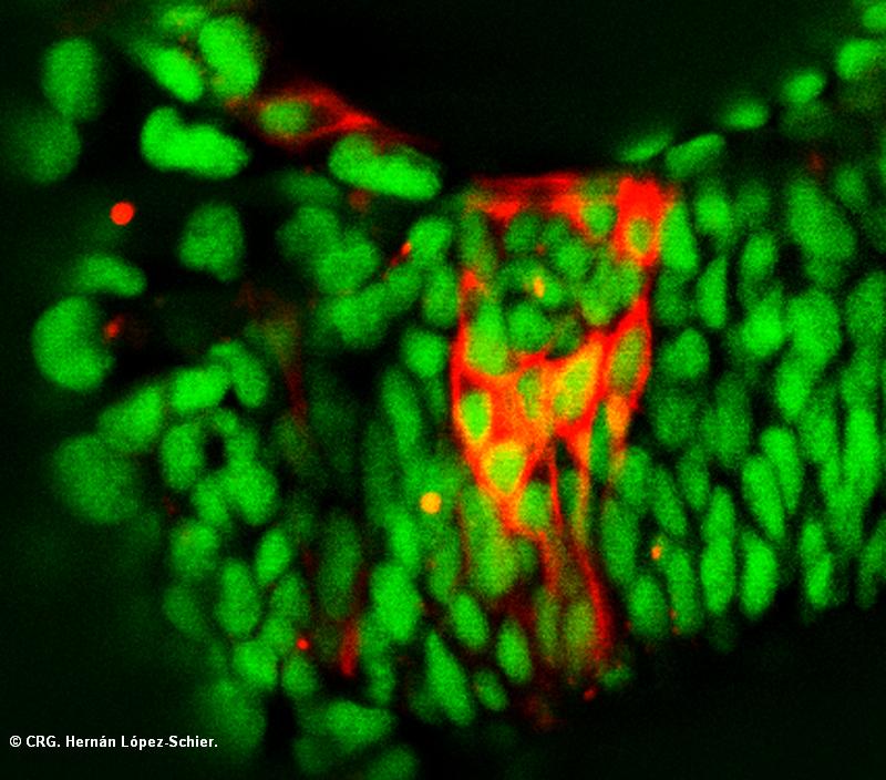
Title: Neurons from zebrafish
Ref: HLS-0025
Author: HERNÁN LÓPEZ-SCHIER
Size: 800 x 802
Description: Transient expression of red fluorescent protein in neurons (red) on a H2A-GFP transgenic line from Jose Campos-Ortega (Cologne, Germany)
Explore CRG Images
The aim of the CRG photo gallery is to provide an overview of our scientists’ research activities through the images generated in the course of their research projects.
All our images are available on demand in digital form with higher quality for for-profit and any other uses. For any of these requests please contact us.
Title: Epitelial cells
Ref: HLS-0024
Author: HERNÁN LÓPEZ-SCHIER
Size: 800 x 704
Description: Transient expression of red fluorescent protein in epithelial cells (red) on a H2A-GFP transgenic line from Jose Campos-Ortega (Cologne, Germany)
Explore CRG Images
The aim of the CRG photo gallery is to provide an overview of our scientists’ research activities through the images generated in the course of their research projects.
All our images are available on demand in digital form with higher quality for for-profit and any other uses. For any of these requests please contact us.
Title: Lateral line from zebrafish
Ref: HLS-0023
Author: HERNÁN LÓPEZ-SCHIER
Size: 800 x 306
Description: Labeling of a lateral line organ of the zebrafish with the antibody HCS1 (red) and Gem25.2 (green) from Teresa Nicolson (USHU, Oregon, USA)
Explore CRG Images
The aim of the CRG photo gallery is to provide an overview of our scientists’ research activities through the images generated in the course of their research projects.
All our images are available on demand in digital form with higher quality for for-profit and any other uses. For any of these requests please contact us.
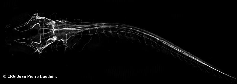
Title: Zebrafish larva neuronal system
Ref: HLS-0021
Author: JEAN-PIERRE BAUDOIN
Size: 800 x 284
Description: Labeling of a zebrafish larva with the antibody mAb 3A10 (neuronal specific)
Explore CRG Images
The aim of the CRG photo gallery is to provide an overview of our scientists’ research activities through the images generated in the course of their research projects.
All our images are available on demand in digital form with higher quality for for-profit and any other uses. For any of these requests please contact us.

Title: Neuromast from trangenic zebrafish
Ref: HLS-0019
Author: HERNÁN LÓPEZ-SCHIER
Size: 800 x 215
Description: Labelling of a neuromast from a transgenic zebrafish for SqET20 (V. Korzh) with an antibody to Caludin b (red) and HCS1 (blue, Jeff Corwin, Virginia). Published at Wibowo et al. 2011
Explore CRG Images
The aim of the CRG photo gallery is to provide an overview of our scientists’ research activities through the images generated in the course of their research projects.
All our images are available on demand in digital form with higher quality for for-profit and any other uses. For any of these requests please contact us.

Title: Neuromast from trangenic zebrafish
Ref: HLS-0018
Author: HERNÁN LÓPEZ-SCHIER
Size: 800 x 800
Description: Labelling of a neuromast from a transgenic zebrafish for SqET20 (V. Korzh) with an antibody to Caludin b (red) and HCS1 (blue, Jeff Corwin, Virginia). Published at Wibowo et al. 2011
Explore CRG Images
The aim of the CRG photo gallery is to provide an overview of our scientists’ research activities through the images generated in the course of their research projects.
All our images are available on demand in digital form with higher quality for for-profit and any other uses. For any of these requests please contact us.