Sensory Cell Biology and Organogenesis
Explore CRG Images
The aim of the CRG photo gallery is to provide an overview of our scientists’ research activities through the images generated in the course of their research projects.
All our images are available on demand in digital form with higher quality for for-profit and any other uses. For any of these requests please contact us.
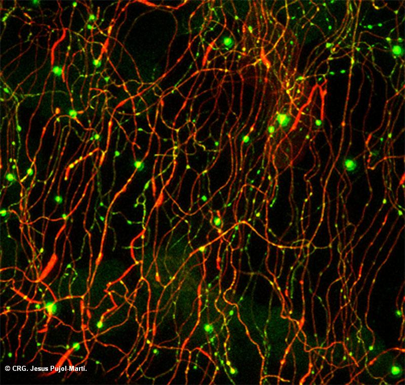
Title: Fireflies in a neuronal jungle
Ref: HLS-0042
Author: Jesús Pujol-Martí
Size: 800 x 762
Description: Peripheral axons of somatosensory neurons that arborize within the skin of a zebrafish larva are labeled in red (Tdtomato). Synaptic boutons are labeled in green (GFP). Axonal arbors shape an intrincate pattern -the jungle- where synapses -the fireflies- pose for our delight.
Explore CRG Images
The aim of the CRG photo gallery is to provide an overview of our scientists’ research activities through the images generated in the course of their research projects.
All our images are available on demand in digital form with higher quality for for-profit and any other uses. For any of these requests please contact us.
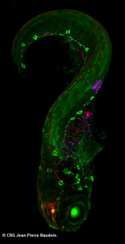
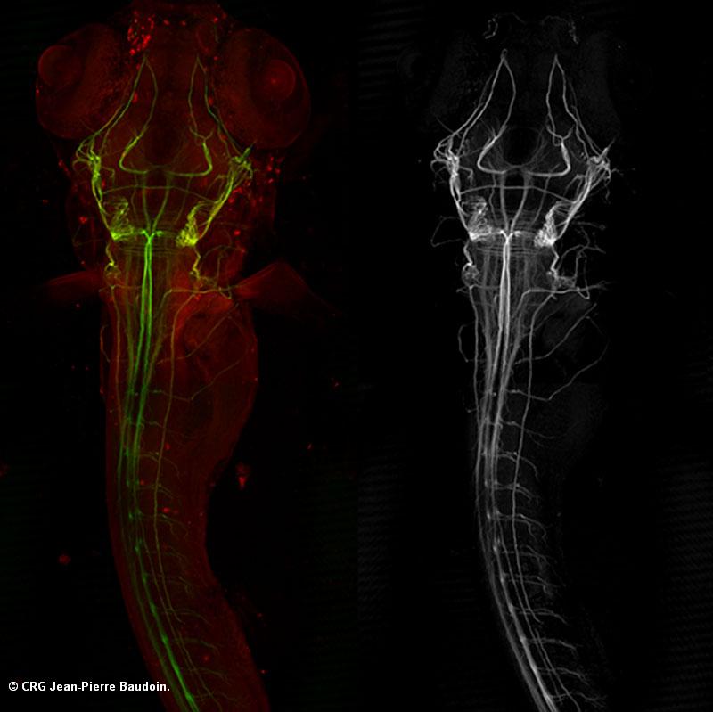
Title: Zebrafish transgenic lines
Ref: HLS-0038
Author: JEAN PIERRE BAUDOIN
Size: 411 x 800
Description: ET20 zebrafish transgenic lines treated with cyclopamine and injected with Magenta dextran
Explore CRG Images
The aim of the CRG photo gallery is to provide an overview of our scientists’ research activities through the images generated in the course of their research projects.
All our images are available on demand in digital form with higher quality for for-profit and any other uses. For any of these requests please contact us.
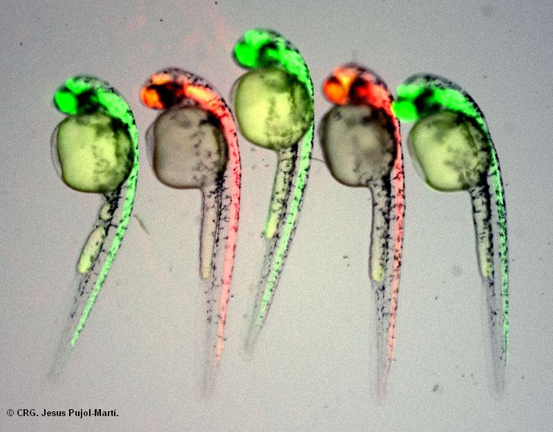
Title: Human-induced diversity in zebrafish embryos
Ref: HLS-0043
Author: Jesús Pujol-Martí
Size: 800 x 626
Description: Kaede is a photoconvertible fluorescent protein. When exposed to UV light, Kaede irreversibly switches from green to red. All the zebrafish embryos shown in this image were at some point Green-Kaede. Two of them, however, were transformed into Red-Kaede by a short exposure to UV light.
Explore CRG Images
The aim of the CRG photo gallery is to provide an overview of our scientists’ research activities through the images generated in the course of their research projects.
All our images are available on demand in digital form with higher quality for for-profit and any other uses. For any of these requests please contact us.
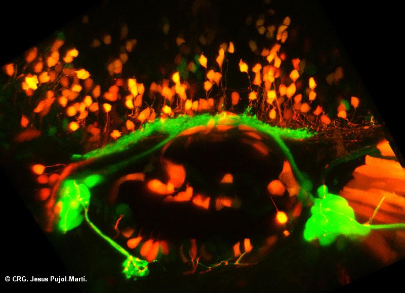
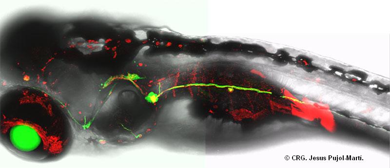
Title: Lateral line sensory neurons and their central targets
Ref: HLS-0041
Author: Jesús Pujol-Martí
Size: 800 x 580
Description: Information captured by the lateral-line sensory system is conveyed to the brain through sensory neurons. There, these neurons make synapses with central neurons that further process the information from the external world. Lateral-line sensory neurons are shown in green (GFP). Their cell bodies form two ganglia (round structures)and their central axons form a bundle that runs rostrocaudally (left-right). Central axons contact the dendrites of central neurons, shown in orange (GFP and Cherry).
Explore CRG Images
The aim of the CRG photo gallery is to provide an overview of our scientists’ research activities through the images generated in the course of their research projects.
All our images are available on demand in digital form with higher quality for for-profit and any other uses. For any of these requests please contact us.

Title: Old and young neurons in the lateral-line ganglion
Ref: HLS-0040
Author: Jesús Pujol-Martí
Size: 800 x 710
Description: Information captured by the lateral-line sensory system is relayed to the brain through sensory neurons, whose cell bodies form a ganglion. By combining transgenics lines that express fluorescent markers at different stages of neuronal development, we can reveal the relative age of neurons. Old neurons(Cherry)occupy the dorsal region of the ganglion whereas young neurons(GFP)occupy the ventral region. A single neuron was back-filled with Alexa 647 Fluor-dextran (in blue).
Explore CRG Images
The aim of the CRG photo gallery is to provide an overview of our scientists’ research activities through the images generated in the course of their research projects.
All our images are available on demand in digital form with higher quality for for-profit and any other uses. For any of these requests please contact us.
Title: Lateral line sensory neurons in the zebrafish larva
Ref: HLS-0039
Author: Jesús Pujol-Martí
Size: 800 x 346
Description: Fish sense water movements in their surroundings by means of the lateral-line sensory system. Information captured by the lateral-line sensory organs, the neuromasts, is relayed to the brain through the lateral-line sensory neurons, highlighted in green (GFP) in the HGn39D transgenic larva of the image. Moreover, a subset of these neurons were labeled by red-fluorescent (rhodamine dextran) injection into a single neuromast. The lens of the eye is also GFP positive in this fish.
Explore CRG Images
The aim of the CRG photo gallery is to provide an overview of our scientists’ research activities through the images generated in the course of their research projects.
All our images are available on demand in digital form with higher quality for for-profit and any other uses. For any of these requests please contact us.
Title: Zebrafish larva neuronal system
Ref: HLS-0037
Author: JEAN PIERRE BAUDOIN
Size: 800 x 799
Description: Labeling of a zebrafish larva with the antibody mAb 3A10 (neuronal specific)
Explore CRG Images
The aim of the CRG photo gallery is to provide an overview of our scientists’ research activities through the images generated in the course of their research projects.
All our images are available on demand in digital form with higher quality for for-profit and any other uses. For any of these requests please contact us.
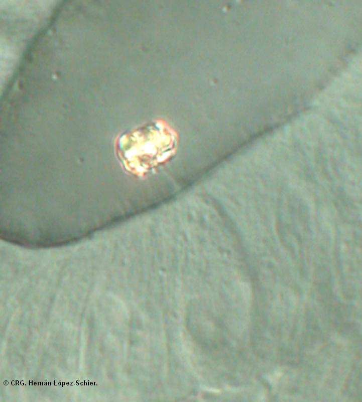
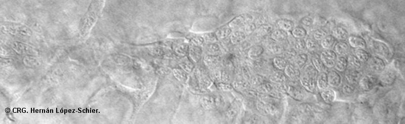
Title: Zebrafish ear with otolith
Ref: HLS-0034
Author: HERNÁN LÓPEZ-SCHIER
Size: 721 x 800
Description: DIC image of a segment of the zebrafish ear showing an otolith
Explore CRG Images
The aim of the CRG photo gallery is to provide an overview of our scientists’ research activities through the images generated in the course of their research projects.
All our images are available on demand in digital form with higher quality for for-profit and any other uses. For any of these requests please contact us.
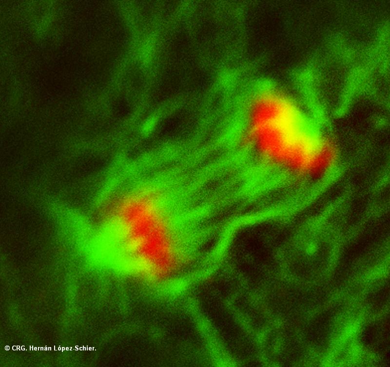
Title: Mitotic spindle with chromosomes
Ref: HLS-0033
Author: HERNÁN LÓPEZ-SCHIER
Size: 800 x 753
Description: Mitotic spindle (green) and chomosomes (red) labelled with antibodies to tubulin (green) and phospho-histone H3 (red)
Explore CRG Images
The aim of the CRG photo gallery is to provide an overview of our scientists’ research activities through the images generated in the course of their research projects.
All our images are available on demand in digital form with higher quality for for-profit and any other uses. For any of these requests please contact us.