Epithelial Homeostasis and Cancer
Explore CRG Images
The aim of the CRG photo gallery is to provide an overview of our scientists’ research activities through the images generated in the course of their research projects.
All our images are available on demand in digital form with higher quality for for-profit and any other uses. For any of these requests please contact us.
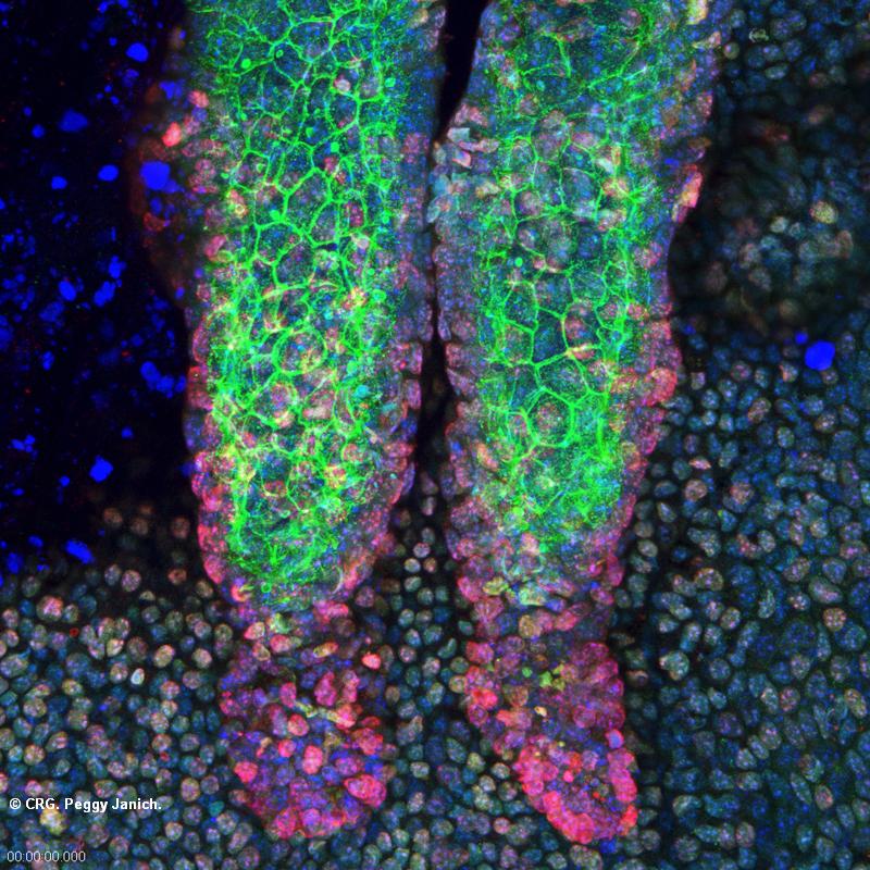
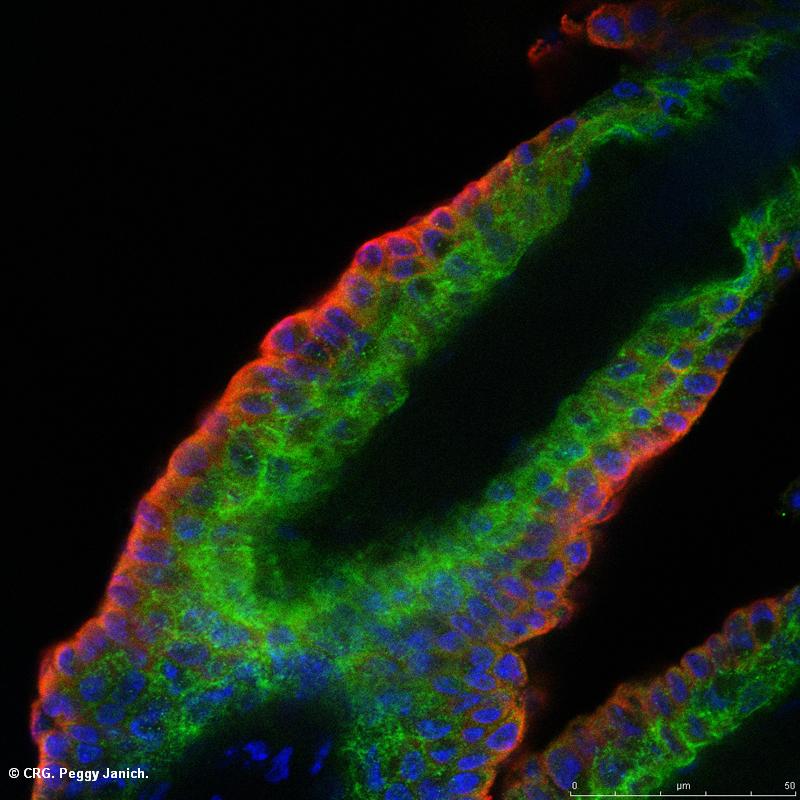
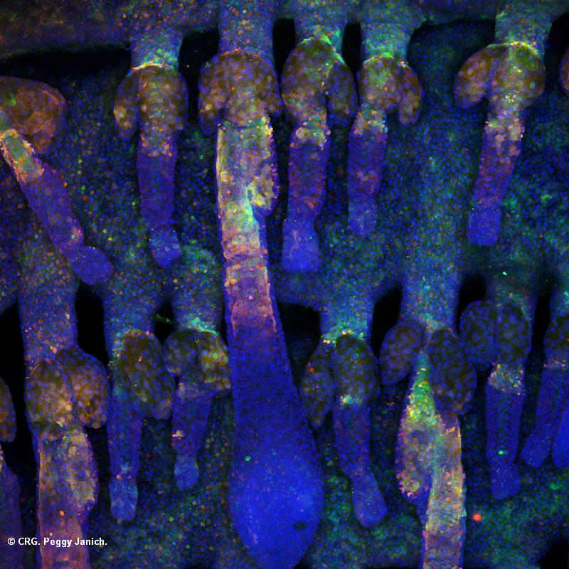
Title: Mouse tail skin 3D reconstruction
Ref: SAB-0017
Author: PEGGY JANICH
Size: 800 x 800
Description: Mouse tail epidermis stained for the protein CLOCK (green), the protein BMAL1 (red) and cell nuclei (DAPI, blue)
Explore CRG Images
The aim of the CRG photo gallery is to provide an overview of our scientists’ research activities through the images generated in the course of their research projects.
All our images are available on demand in digital form with higher quality for for-profit and any other uses. For any of these requests please contact us.
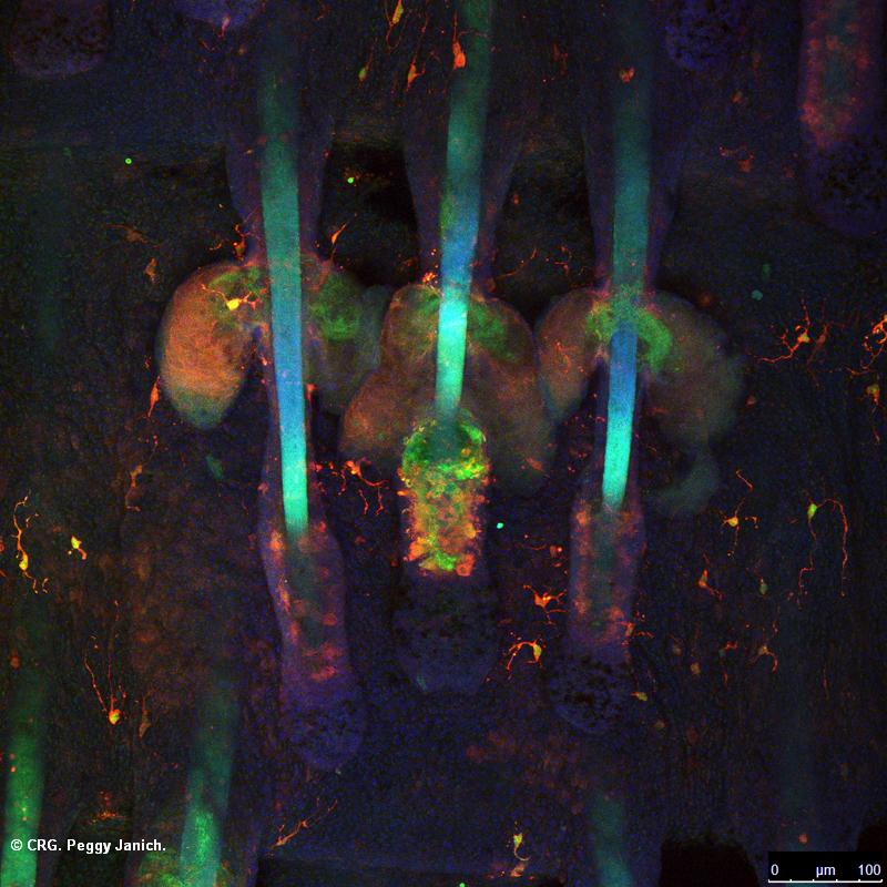
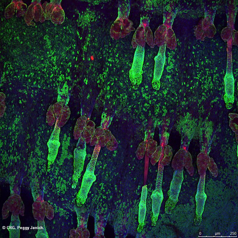
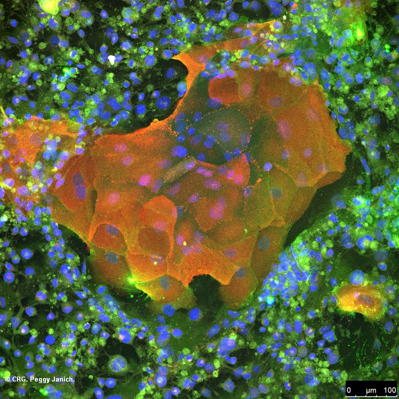
Title: Mouse tail skin 3D reconstruction
Ref: SAB-0016
Author: PEGGY JANICH
Size: 800 x 800
Description: Mouse tail epidermis of a transgenic mouse expressing the green fluorescent protein (GFP, green) under a gene specific promoter was stained for GFP (anti-GFP antibody, red) and cell nuclei (DAPI, blue)
Explore CRG Images
The aim of the CRG photo gallery is to provide an overview of our scientists’ research activities through the images generated in the course of their research projects.
All our images are available on demand in digital form with higher quality for for-profit and any other uses. For any of these requests please contact us.
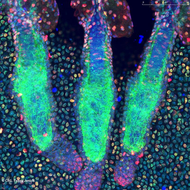
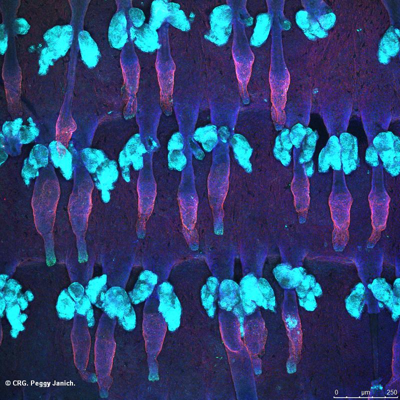
Title: Mouse tail skin 3D reconstruction
Ref: SAB-0015
Author: PEGGY JANICH
Size: 800 x 800
Description: Mouse tail epidermis stained for the protein CLOCK (green), the protein BMAL1 (red) and cell nuclei (DAPI, blue)
Explore CRG Images
The aim of the CRG photo gallery is to provide an overview of our scientists’ research activities through the images generated in the course of their research projects.
All our images are available on demand in digital form with higher quality for for-profit and any other uses. For any of these requests please contact us.
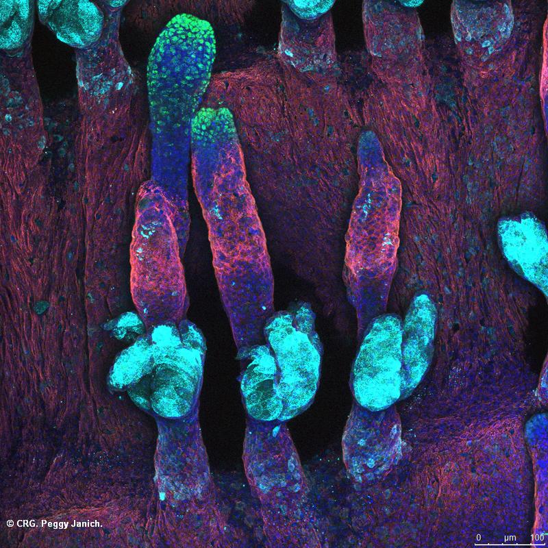
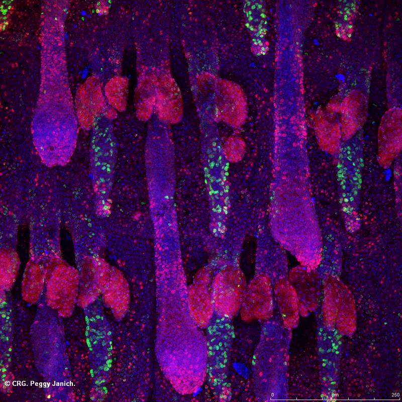
Title: Mouse tail skin 3D reconstruction
Ref: SAB-0014
Author: PEGGY JANICH
Size: 800 x 800
Description: Mouse tail epidermis stained for the protein CDP (green), alpha6 integrin (red), beta-galactosidase (cyan) and cell nuclei (DAPI, blue)
Explore CRG Images
The aim of the CRG photo gallery is to provide an overview of our scientists’ research activities through the images generated in the course of their research projects.
All our images are available on demand in digital form with higher quality for for-profit and any other uses. For any of these requests please contact us.
Title: Mouse tail skin 3D reconstruction
Ref: SAB-0013
Author: PEGGY JANICH
Size: 800 x 800
Description: Mouse tail epidermis stained for the protein CLOCK (green), Keratin 15 (red), and cell nuclei (DAPI, blue)
Explore CRG Images
The aim of the CRG photo gallery is to provide an overview of our scientists’ research activities through the images generated in the course of their research projects.
All our images are available on demand in digital form with higher quality for for-profit and any other uses. For any of these requests please contact us.
Title: Mouse tail skin 3D reconstruction
Ref: SAB-0012
Author: PEGGY JANICH
Size: 800 x 800
Description: Mouse tail epidermis stained for Keratin 14 (green), Lrig1 (red), and cell nuclei (DAPI, blue)
Explore CRG Images
The aim of the CRG photo gallery is to provide an overview of our scientists’ research activities through the images generated in the course of their research projects.
All our images are available on demand in digital form with higher quality for for-profit and any other uses. For any of these requests please contact us.
Title: Mouse tail skin 3D reconstruction
Ref: SAB-0011
Author: PEGGY JANICH
Size: 800 x 800
Description: Mouse tail epidermis stained for alpha6 integrin (red), the protein CDP (green), beta-galactosidase (cyan) and cell nuclei (DAPI, blue)
Explore CRG Images
The aim of the CRG photo gallery is to provide an overview of our scientists’ research activities through the images generated in the course of their research projects.
All our images are available on demand in digital form with higher quality for for-profit and any other uses. For any of these requests please contact us.
Title: Mouse tail skin 3D reconstruction
Ref: SAB-0010
Author: PEGGY JANICH
Size: 800 x 800
Description: Mouse tail epidermis stained for a proliferation marker (Ki67, red), label retaining cells (BrdU, green) and cell nuclei (DAPI, blue)
Explore CRG Images
The aim of the CRG photo gallery is to provide an overview of our scientists’ research activities through the images generated in the course of their research projects.
All our images are available on demand in digital form with higher quality for for-profit and any other uses. For any of these requests please contact us.
Title: Mouse tail skin 3D reconstruction
Ref: SAB-0009
Author: PEGGY JANICH
Size: 800 x 800
Description: Mouse tail epidermis of a transgenic mouse expressing the green fluorescent protein (GFP, green) under a gene specific promoter was stained for GFP (anti-GFP antibody, red) and cell nuclei (DAPI, blue)
Explore CRG Images
The aim of the CRG photo gallery is to provide an overview of our scientists’ research activities through the images generated in the course of their research projects.
All our images are available on demand in digital form with higher quality for for-profit and any other uses. For any of these requests please contact us.