Epithelial Homeostasis and Cancer
Explore CRG Images
The aim of the CRG photo gallery is to provide an overview of our scientists’ research activities through the images generated in the course of their research projects.
All our images are available on demand in digital form with higher quality for for-profit and any other uses. For any of these requests please contact us.
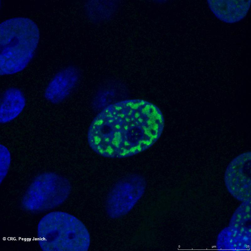
Title: Mouse skin hair follicles
Ref: SAB-0034
Author: PEGGY JANICH
Size: 800 x 800
Description:
-
Explore CRG Images
The aim of the CRG photo gallery is to provide an overview of our scientists’ research activities through the images generated in the course of their research projects.
All our images are available on demand in digital form with higher quality for for-profit and any other uses. For any of these requests please contact us.
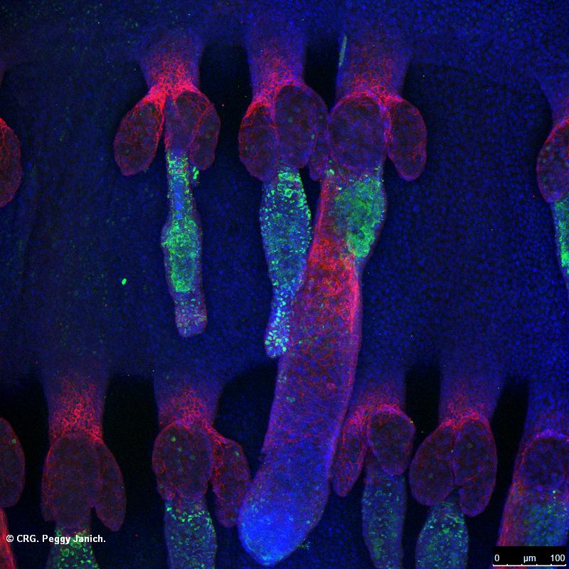
Title: 3D reconstruction of mouse skin
Ref: SAB-0032
Author: PEGGY JANICH
Size: 800 x 800
Description:
-
Explore CRG Images
The aim of the CRG photo gallery is to provide an overview of our scientists’ research activities through the images generated in the course of their research projects.
All our images are available on demand in digital form with higher quality for for-profit and any other uses. For any of these requests please contact us.
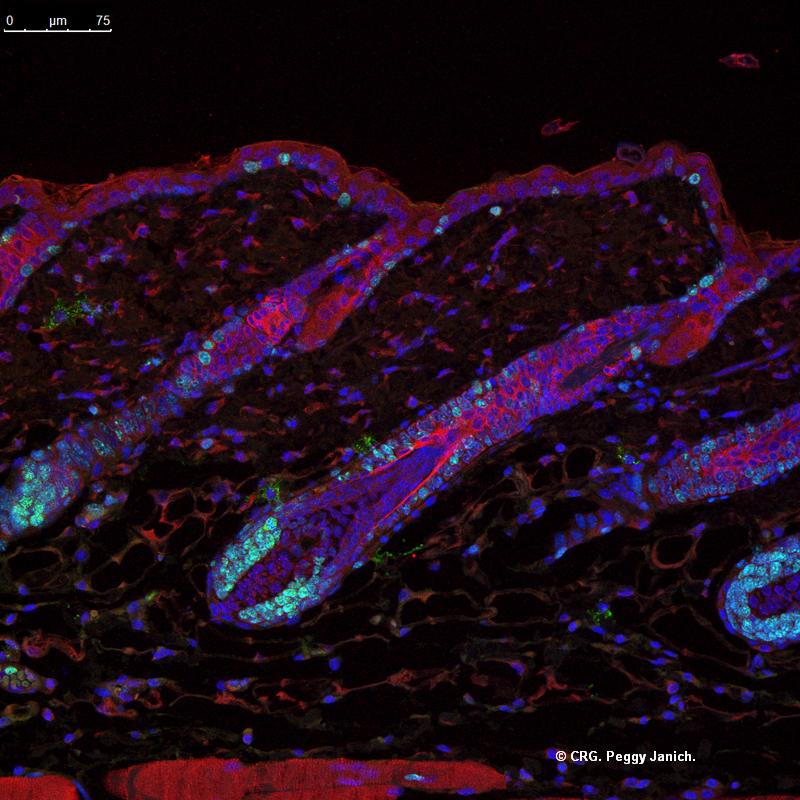
Title: Mouse skin hair follicle section
Ref: SAB-0030
Author: PEGGY JANICH
Size: 800 x 800
Description:
-
Explore CRG Images
The aim of the CRG photo gallery is to provide an overview of our scientists’ research activities through the images generated in the course of their research projects.
All our images are available on demand in digital form with higher quality for for-profit and any other uses. For any of these requests please contact us.

Title: Quiescent cancer stem cell
Ref: SAB-0029
Author: stefania mejetta
Size: 800 x 800
Description: Quiescent cancer stem cell (green) are found in the basal CD44+ population (red) of a human xenograft of squamous cell carcinoma (human tumor cells are blue).
Explore CRG Images
The aim of the CRG photo gallery is to provide an overview of our scientists’ research activities through the images generated in the course of their research projects.
All our images are available on demand in digital form with higher quality for for-profit and any other uses. For any of these requests please contact us.
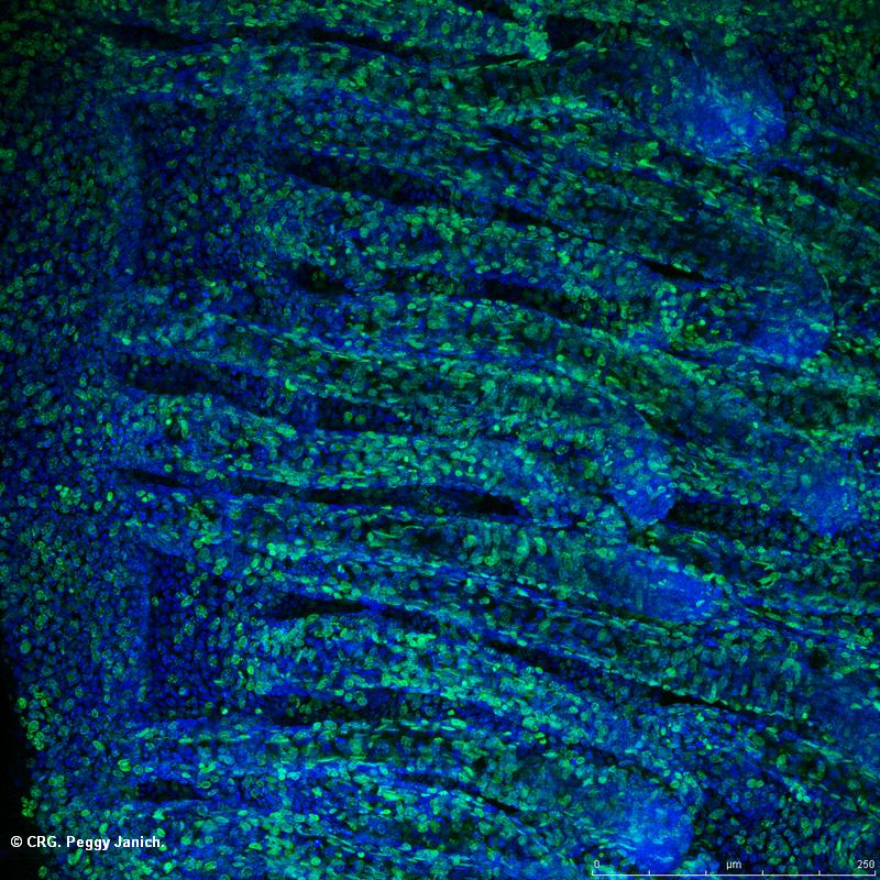
Title: Mouse tail skin 3D reconstruction
Ref: SAB-0024
Author: PEGGY JANICH
Size: 800 x 800
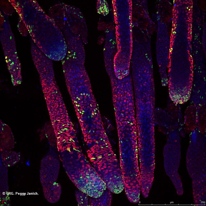
Description: Mouse tail epidermis stained for a proliferation marker (Ki67, green) and cell nuclei (DAPI, blue)
Explore CRG Images
The aim of the CRG photo gallery is to provide an overview of our scientists’ research activities through the images generated in the course of their research projects.
All our images are available on demand in digital form with higher quality for for-profit and any other uses. For any of these requests please contact us.
Title: Mouse tail skin 3D reconstruction
Ref: SAB-0023
Author: PEGGY JANICH
Size: 800 x 800
Description: Mouse tail epidermis stained for the protein CLOCK (red), sebaceous glands (green) and cell nuclei (DAPI, blue)
Explore CRG Images
The aim of the CRG photo gallery is to provide an overview of our scientists’ research activities through the images generated in the course of their research projects.
All our images are available on demand in digital form with higher quality for for-profit and any other uses. For any of these requests please contact us.
Title: Mouse tail skin 3D reconstruction
Ref: SAB-0022
Author: PEGGY JANICH
Size: 800 x 800
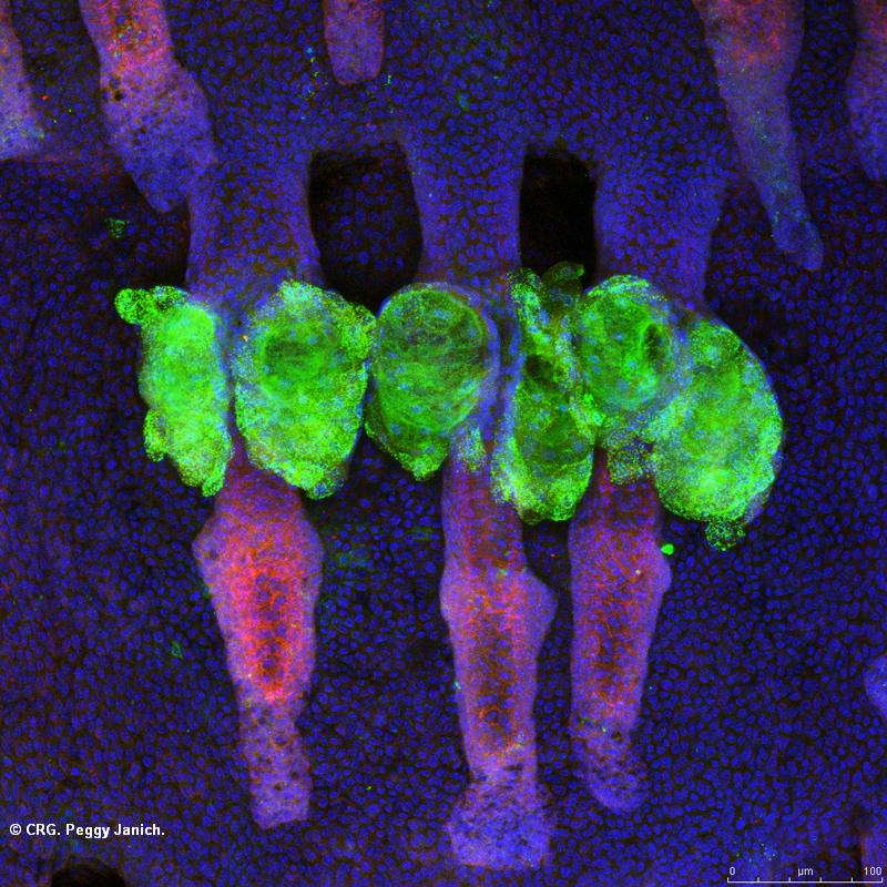
Description: Mouse tail epidermis stained for a proliferation marker (Ki67, green), the protein BMAL1 (red) and cell nuclei (DAPI, blue)
Explore CRG Images
The aim of the CRG photo gallery is to provide an overview of our scientists’ research activities through the images generated in the course of their research projects.
All our images are available on demand in digital form with higher quality for for-profit and any other uses. For any of these requests please contact us.

Title: Mouse tail skin 3D reconstruction
Ref: SAB-0020
Author: PEGGY JANICH
Size: 800 x 800
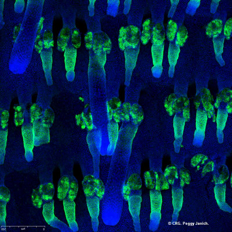
Description: Mouse tail epidermis of a TOPGAL transgenic mouse stained for beta-galactosidase (green) and cell nuclei (DAPI, blue)
Explore CRG Images
The aim of the CRG photo gallery is to provide an overview of our scientists’ research activities through the images generated in the course of their research projects.
All our images are available on demand in digital form with higher quality for for-profit and any other uses. For any of these requests please contact us.
Title: Mouse tail skin 3D reconstruction
Ref: SAB-0019
Author: PEGGY JANICH
Size: 800 x 800
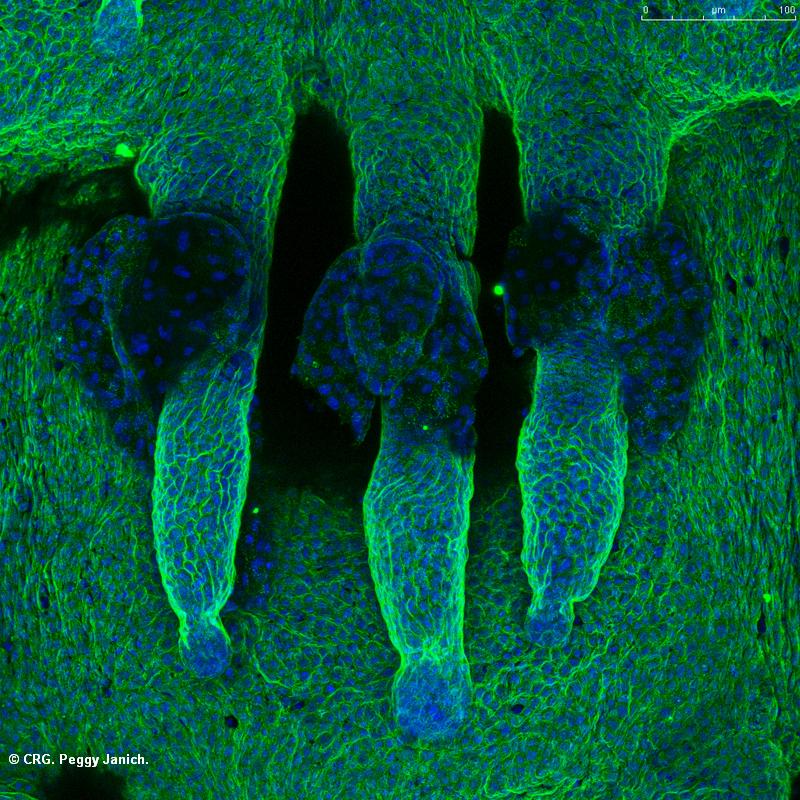
Description: Mouse tail epidermis stained for alpha6-integrin (green) and cell nuclei (DAPI, blue)
Explore CRG Images
The aim of the CRG photo gallery is to provide an overview of our scientists’ research activities through the images generated in the course of their research projects.
All our images are available on demand in digital form with higher quality for for-profit and any other uses. For any of these requests please contact us.