Explore CRG Images
The aim of the CRG photo gallery is to provide an overview of our scientists’ research activities through the images generated in the course of their research projects.
All our images are available on demand in digital form with higher quality for for-profit and any other uses. For any of these requests please contact us.

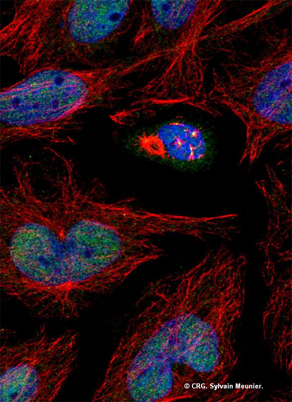
Title: Microtubule regrowth from centrosomes and chromosomes
Ref: IV-0038
Author: Sylvain Meunier
CRG Group: Microtubules Function and Cell Division - Isabelle Vernos
Size: 581 x 800
Description: After total microtubule depolymerization by nocodazole treatment, the drug is washed out and the microtubules (red) start to regrow in the mitotic cell from the centrosomes and the chromosomes (blue). MCRS1, stained in green, is the first specific marker for acentrosomal microtubule assembly, and plays a role in the control of the dynamics of this microtubule subclass.

