Multicellular Systems Biology
Explore CRG Images
The aim of the CRG photo gallery is to provide an overview of our scientists’ research activities through the images generated in the course of their research projects.
All our images are available on demand in digital form with higher quality for for-profit and any other uses. For any of these requests please contact us.
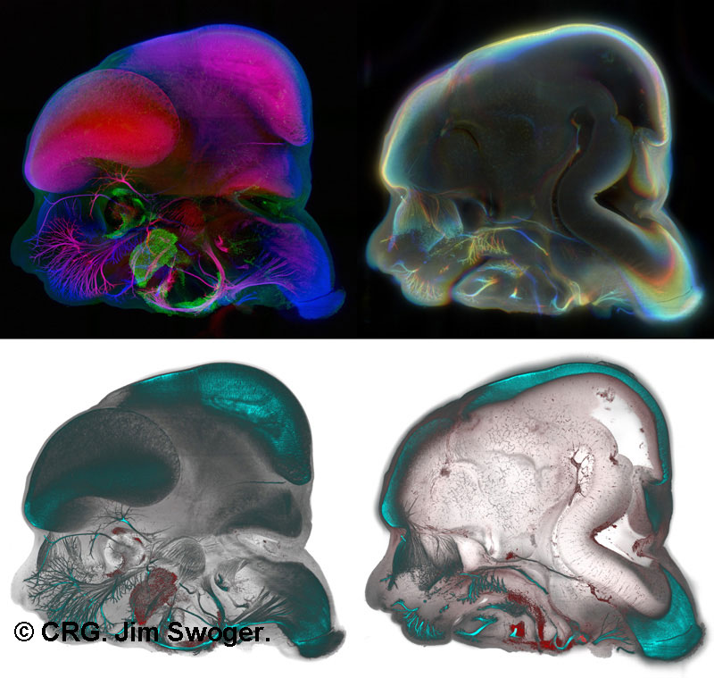
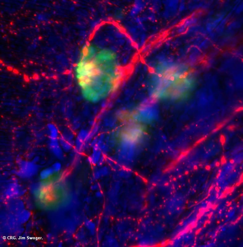
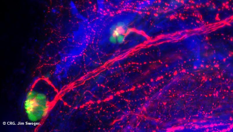
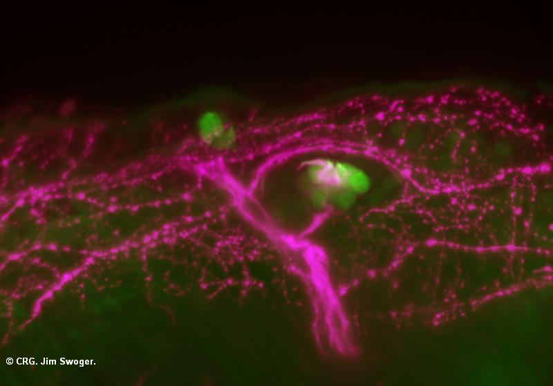
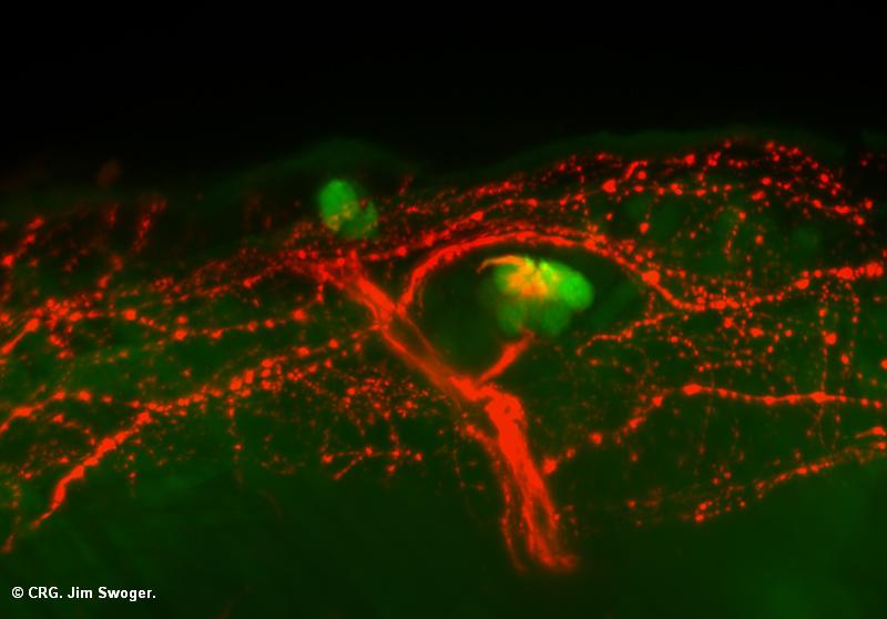
Title: Visualizations of an Embryonic Mouse Head
Ref: JS-0077
Author: Jim Swoger
Size: 800 x 759
Description: An E12.0 mouse embryo was fluorescently labeled to visualize neural (lower images, cyan) and muscle precursor (lower, red) tissue, and a Selective Plane Illumination Microscopy scan was done of the head. Four alternative visualizations of the 3D data set are presented. Left: projections through the left half of the head (thickness 1mm). Right: projections through a virtual section (thickness 0.25mm). Upper left: nerves red-blue depth-coded; muscle precursors green. Upper right: nerves depth-coded. Bottom: alternative representations of the data used for the upper images.
Explore CRG Images
The aim of the CRG photo gallery is to provide an overview of our scientists’ research activities through the images generated in the course of their research projects.
All our images are available on demand in digital form with higher quality for for-profit and any other uses. For any of these requests please contact us.
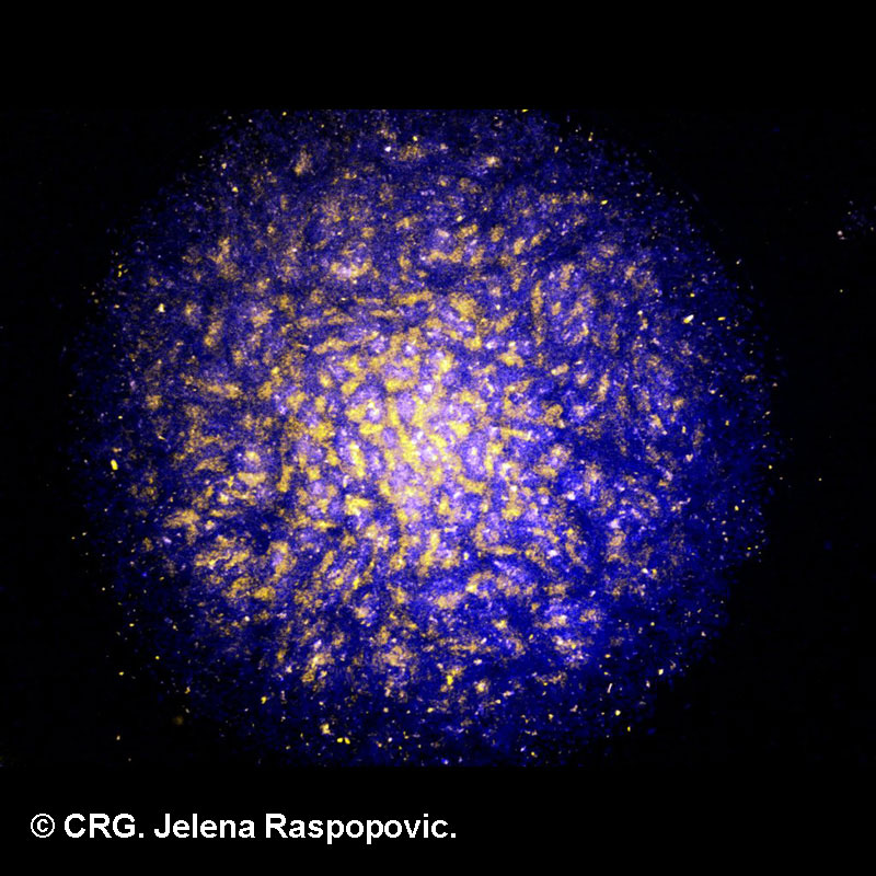
Title: Micromass Galaxy I
Ref: JS-0076
Author: Jelena Raspopovic
Size: 800 x 800
Description: Mesenchymal cells, extracted from a developing mouse limb, form a periodic pattern as seen in this picture. This culture has a particular name of micromass cell culture. In blue you can see cells that will differentiate into bone and cartilage, they express Sox9 transcription factor, and in yellow the ones that will not, they express Bmp2 protein. This pattern arises spontaneously from a homogeneous state of cells.
Explore CRG Images
The aim of the CRG photo gallery is to provide an overview of our scientists’ research activities through the images generated in the course of their research projects.
All our images are available on demand in digital form with higher quality for for-profit and any other uses. For any of these requests please contact us.
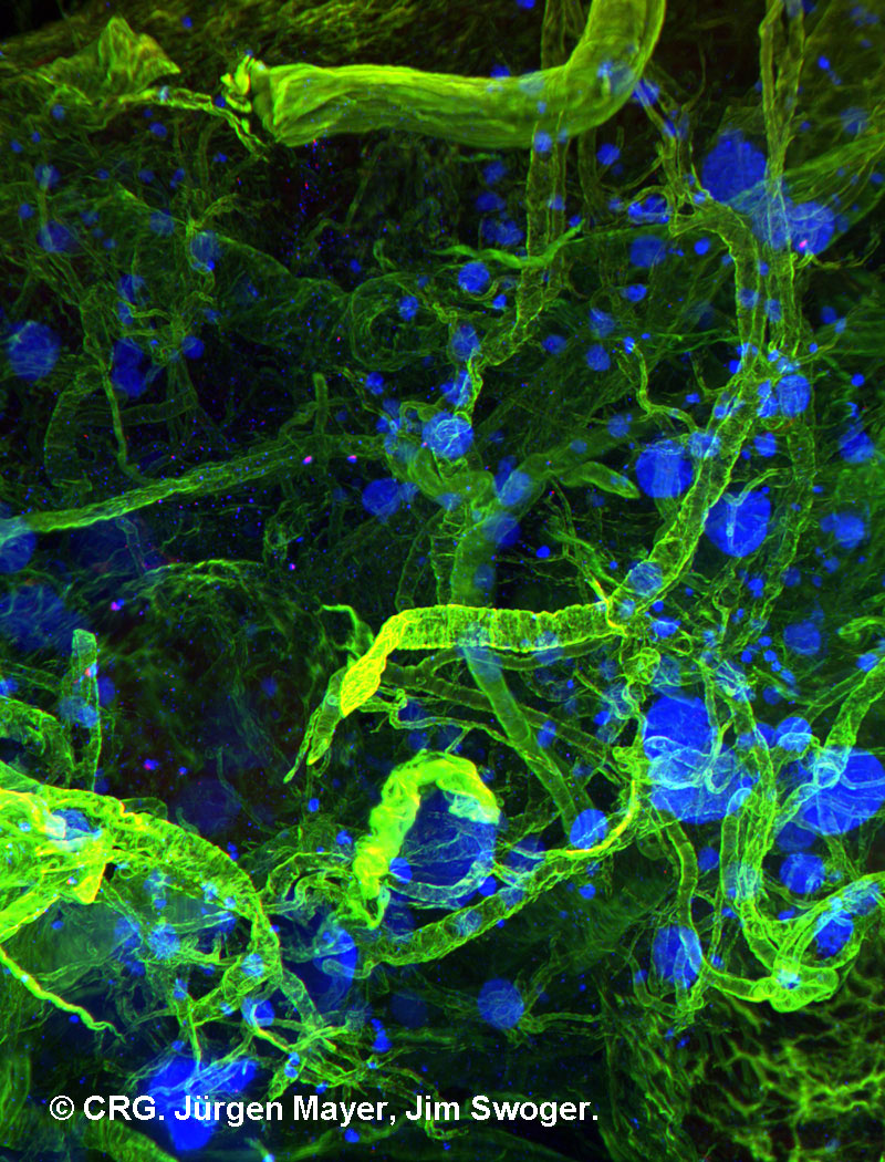
Title: Deep inside the pancreas
Ref: JS-0075
Author: Jürgen Mayer, Jim Swoger
Size: 800 x 1050
Description: Part of a murine pancreas imaged with an experimental light sheet microscope setup. Blood vessels in green, labeled with Asma-Cy3. Insulin producing beta-cells in blue, labeled with IR647 anti-GP. Beta-cells contribute up to 80% of the cells in Islets of Langerhans, the larger blue structures in the image. The original dimensions of the pancreatic tissue are 1.3 x 1.7 x 7.2 mm. The image is a maximum value projection of 1441 slices, so the spacing is 5 µm.
Explore CRG Images
The aim of the CRG photo gallery is to provide an overview of our scientists’ research activities through the images generated in the course of their research projects.
All our images are available on demand in digital form with higher quality for for-profit and any other uses. For any of these requests please contact us.
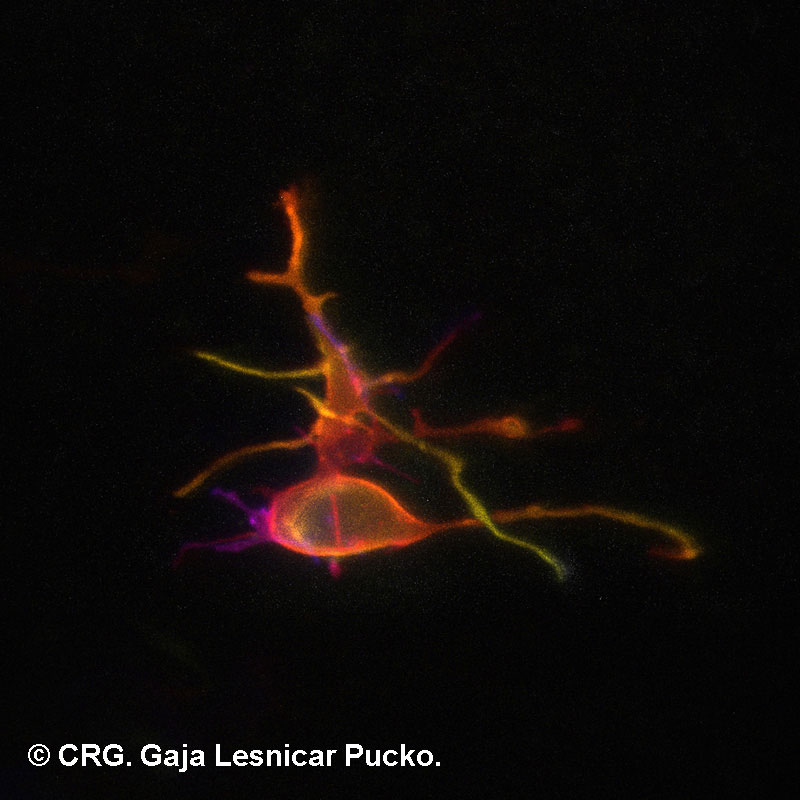
Title: Burning Cell
Ref: JS-0074
Author: Gaja Lesnicar Pucko
Size: 800 x 800
Description: Mesenchymal cell expressing membrane targeted EGFP. This image was taken with a novel in ovo microscopy approach. We electroporated young chick embryo with pCAGGS-gpiEGFP vector to obtain a salt and pepper distribution of labelled cells. The egg was sealed with Teflon membrane that allowed gas exchange and embryo visualization and placed under the CLSM microscope. A 30 microns thick z-stack of 0,5 micron steps was taken. Z-stack was color coded for depth and maximum projected in Fiji. I like the false solitude of this cell – in reality it is surrounded by a multitude of unlabelled cells.
Explore CRG Images
The aim of the CRG photo gallery is to provide an overview of our scientists’ research activities through the images generated in the course of their research projects.
All our images are available on demand in digital form with higher quality for for-profit and any other uses. For any of these requests please contact us.
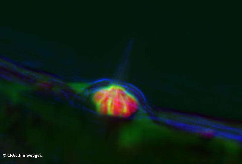
Title: Hair cells in a zebrafish
Ref: JS-0052
Author: Jim Swoger
Size: 786 x 800
Description: A SPIM image of sensory hair cells in the developing zebrafish.
Explore CRG Images
The aim of the CRG photo gallery is to provide an overview of our scientists’ research activities through the images generated in the course of their research projects.
All our images are available on demand in digital form with higher quality for for-profit and any other uses. For any of these requests please contact us.
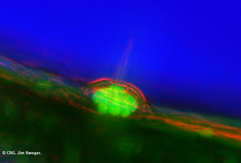
Title: Hair cells in a zebrafish
Ref: JS-0051
Author: Jim Swoger
Size: 800 x 453
Description: A SPIM image of sensory hair cells in the developing zebrafish.
Explore CRG Images
The aim of the CRG photo gallery is to provide an overview of our scientists’ research activities through the images generated in the course of their research projects.
All our images are available on demand in digital form with higher quality for for-profit and any other uses. For any of these requests please contact us.
Title: Hair cells in a zebrafish
Ref: JS-0050
Author: Jim Swoger
Size: 800 x 558
Description: A SPIM image of sensory hair cells in the developing zebrafish.
Explore CRG Images
The aim of the CRG photo gallery is to provide an overview of our scientists’ research activities through the images generated in the course of their research projects.
All our images are available on demand in digital form with higher quality for for-profit and any other uses. For any of these requests please contact us.
Title: Hair cells in a zebrafish
Ref: JS-0049
Author: Jim Swoger
Size: 800 x 558
Description: A SPIM image of sensory hair cells in the developing zebrafish.
Explore CRG Images
The aim of the CRG photo gallery is to provide an overview of our scientists’ research activities through the images generated in the course of their research projects.
All our images are available on demand in digital form with higher quality for for-profit and any other uses. For any of these requests please contact us.
Title: Hair cells in a zebrafish
Ref: JS-0048
Author: Jim Swoger
Size: 800 x 541
Description: A SPIM image of sensory hair cells in the developing zebrafish.
Explore CRG Images
The aim of the CRG photo gallery is to provide an overview of our scientists’ research activities through the images generated in the course of their research projects.
All our images are available on demand in digital form with higher quality for for-profit and any other uses. For any of these requests please contact us.