Microtubules Function and Cell Division
Explore CRG Images
The aim of the CRG photo gallery is to provide an overview of our scientists’ research activities through the images generated in the course of their research projects.
All our images are available on demand in digital form with higher quality for for-profit and any other uses. For any of these requests please contact us.
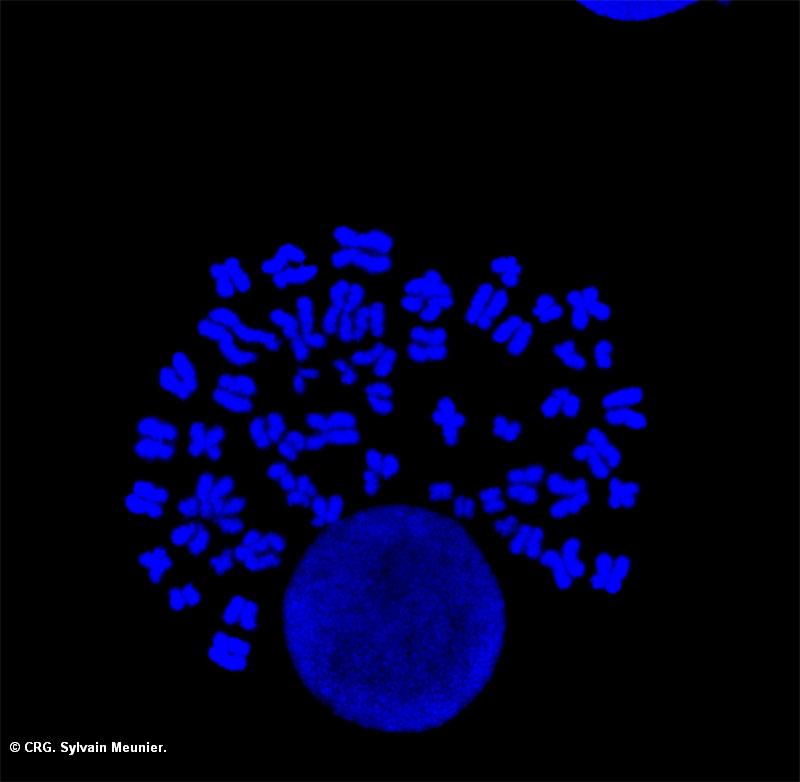
Title: Bunch of chromosomes
Ref: IV-0039
Author: Sylvain Meunier
Size: 800 x 782
Description: Mitotic chromosome forming a bouquet around an intact interphasic nucleus
Explore CRG Images
The aim of the CRG photo gallery is to provide an overview of our scientists’ research activities through the images generated in the course of their research projects.
All our images are available on demand in digital form with higher quality for for-profit and any other uses. For any of these requests please contact us.
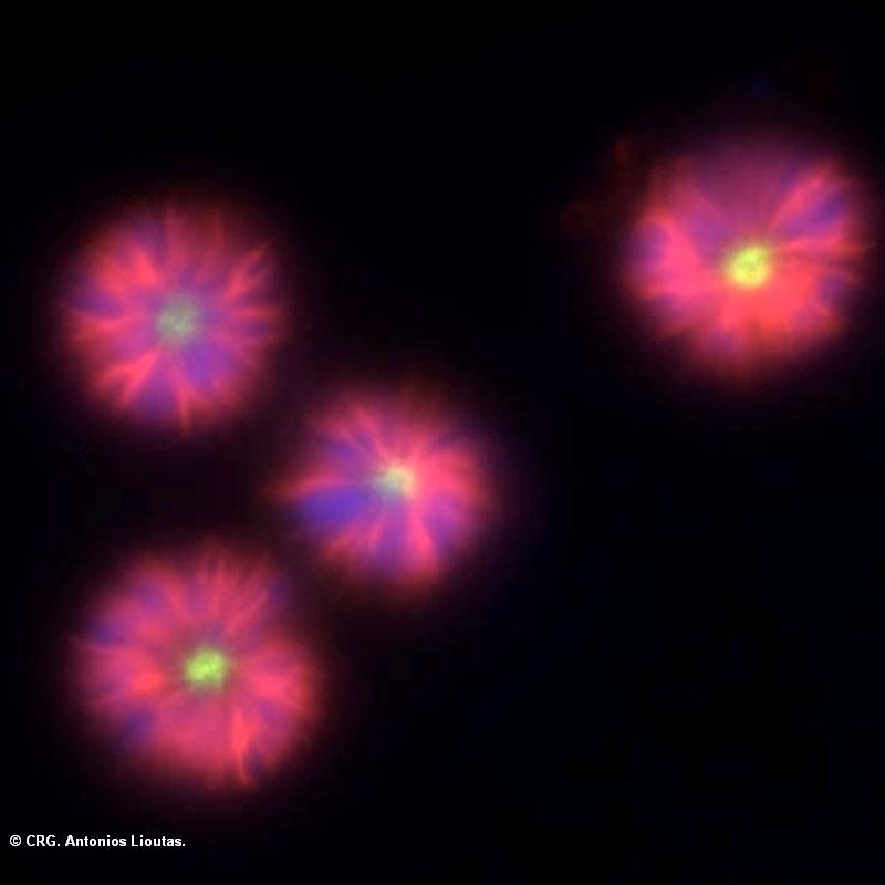
Title: Monopolar spindles
Ref: IV-0018
Author: ANTONIOS LIOUTAS
Size: 800 x 800
Description: Immunofluorescence image of monopolar spindles formed in cells treated with an inhibitor (STLC) for the motor protein Eg5 (kinesin-5). Microtubules are shown in red, chromosomes in blue and Phospho-Aurora A in green.
Explore CRG Images
The aim of the CRG photo gallery is to provide an overview of our scientists’ research activities through the images generated in the course of their research projects.
All our images are available on demand in digital form with higher quality for for-profit and any other uses. For any of these requests please contact us.
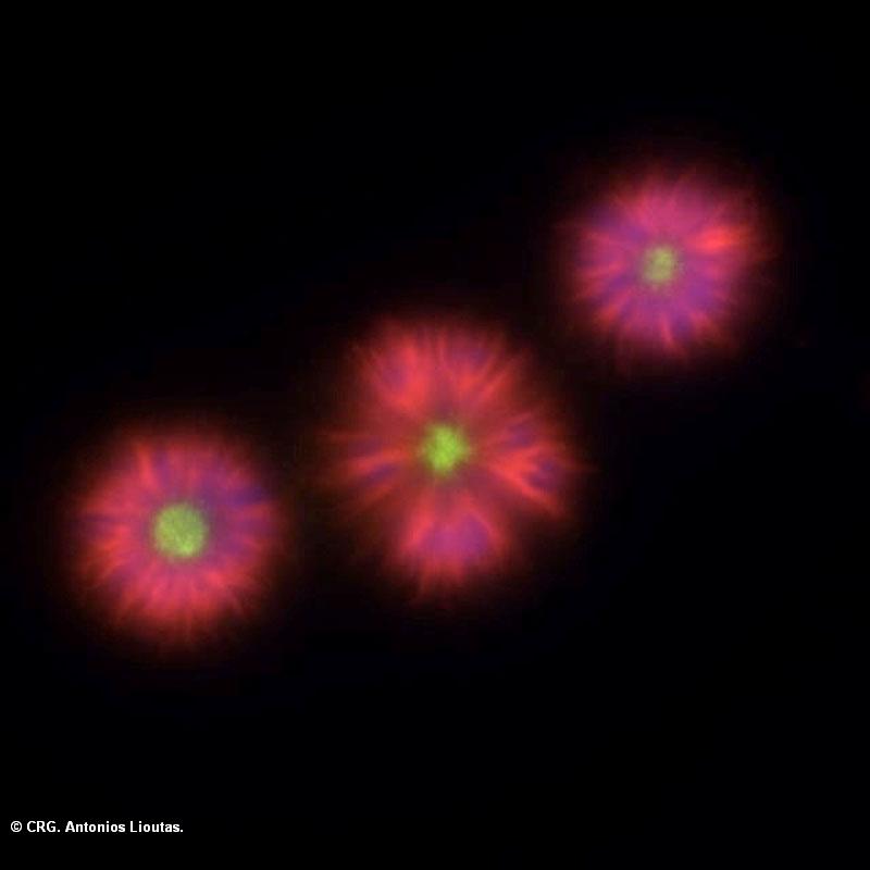
Title: Monopolar spindles
Ref: IV-0017
Author: ANTONIOS LIOUTAS
Size: 800 x 800
Description: Immunofluorescence image of monopolar spindles formed in cells treated with an inhibitor (STLC) for the motor protein Eg5 (kinesin-5). Microtubules are shown in red, chromosomes in blue and Phospho-Aurora A in green.
Explore CRG Images
The aim of the CRG photo gallery is to provide an overview of our scientists’ research activities through the images generated in the course of their research projects.
All our images are available on demand in digital form with higher quality for for-profit and any other uses. For any of these requests please contact us.
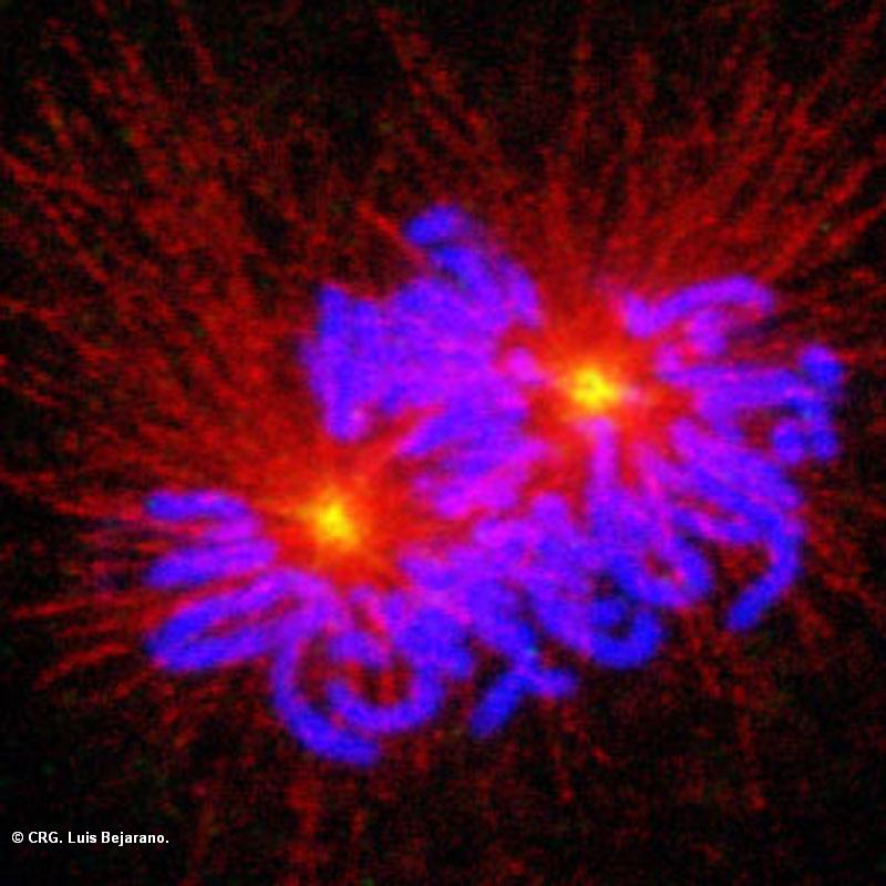
Title: Xenopus cell in prometaphase
Ref: IV-0016
Author: Luis Bejarano
Size: 800 x 800
Description: Microtubules are visualized in red and chromosomes in blue. The localization of the mitotic kinase Aurora A at the center of the two microtubule asters of the forming spindle is shown in green (yellow in the overlay).
Explore CRG Images
The aim of the CRG photo gallery is to provide an overview of our scientists’ research activities through the images generated in the course of their research projects.
All our images are available on demand in digital form with higher quality for for-profit and any other uses. For any of these requests please contact us.
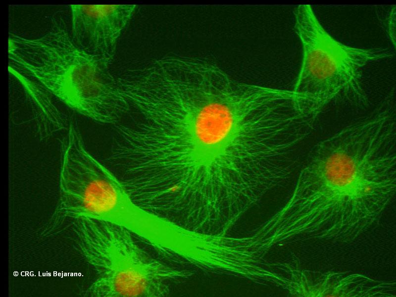
Title: Xenopus cells in culture
Ref: IV-0015
Author: Luis Bejarano
Size: 800 x 600
Description: Immunofluorescent image of interphase Xenopus cells (XL177) in culture. Microtubules are visualized in green and the DNA in red.
Explore CRG Images
The aim of the CRG photo gallery is to provide an overview of our scientists’ research activities through the images generated in the course of their research projects.
All our images are available on demand in digital form with higher quality for for-profit and any other uses. For any of these requests please contact us.

Title: Xenopus cell in prometaphase
Ref: IV-0014
Author: Luis Bejarano
Size: 800 x 800
Description: Microtubules are visualized in red and chromosomes in blue. The localization of the mitotic kinase Aurora A at the spindle poles is shown in green (yellow in the overlay). Image obtained with the Leica SP5 confocal microscope.
Explore CRG Images
The aim of the CRG photo gallery is to provide an overview of our scientists’ research activities through the images generated in the course of their research projects.
All our images are available on demand in digital form with higher quality for for-profit and any other uses. For any of these requests please contact us.
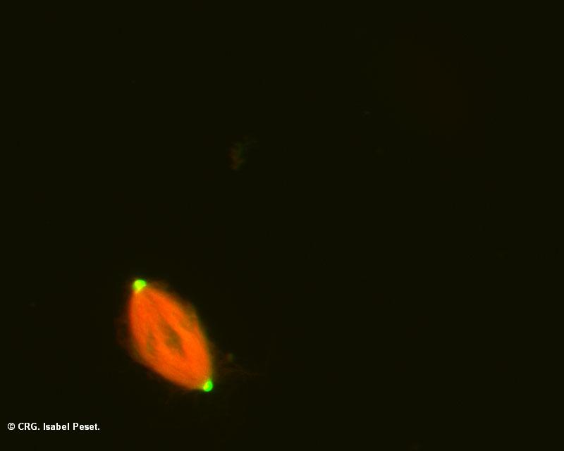
Title: Spindle assembled in Xenopus egg extract
Ref: IV-0006
Author: Isabel Peset
Size: 800 x 640
Description: Microtubules are shown in red and DNA in blue. The signal in green at the spindle poles correspond to GFP tagged TACC domain of the protein Maskin that was added to the extract.
Explore CRG Images
The aim of the CRG photo gallery is to provide an overview of our scientists’ research activities through the images generated in the course of their research projects.
All our images are available on demand in digital form with higher quality for for-profit and any other uses. For any of these requests please contact us.
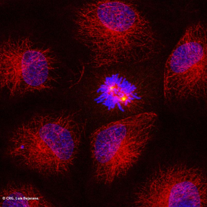
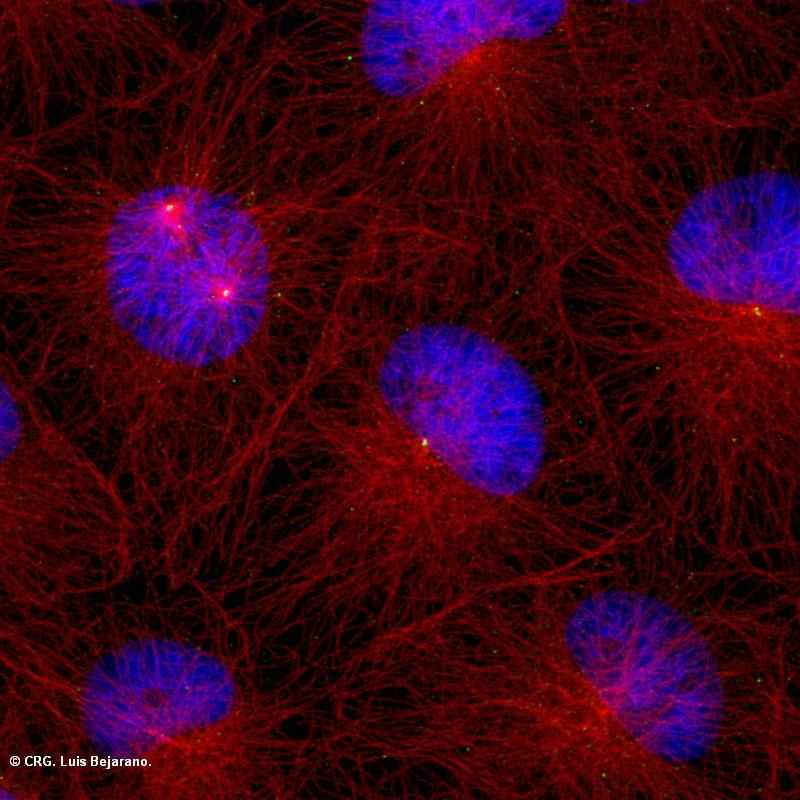
Title: Xenopus cell in prometaphase
Ref: IV-0004
Author: Luis Bejarano
Size: 800 x 800
Description: Microtubules are visualized in red and chromosomes in blue. The localization of the mitotic kinase Aurora A at the spindle poles is shown in green (yellow in the overlay). Surrounding cells are in interphase.
Explore CRG Images
The aim of the CRG photo gallery is to provide an overview of our scientists’ research activities through the images generated in the course of their research projects.
All our images are available on demand in digital form with higher quality for for-profit and any other uses. For any of these requests please contact us.
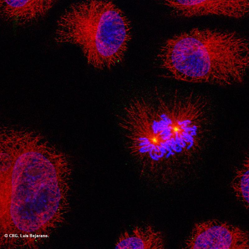
Title: Xenopus cell in prometaphase
Ref: IV-0003
Author: Luis Bejarano
Size: 800 x 800
Description: Microtubules are visualized in red and chromosomes in blue. The localization of the mitotic kinase Aurora A at the center of the two microtubule asters of the forming spindle is shown in green (yellow in the overlay). Surrounding cells are in interphase.
Explore CRG Images
The aim of the CRG photo gallery is to provide an overview of our scientists’ research activities through the images generated in the course of their research projects.
All our images are available on demand in digital form with higher quality for for-profit and any other uses. For any of these requests please contact us.
Title: Xenopus cell in prophase
Ref: IV-0002
Author: Luis Bejarano
Size: 800 x 800
Description: Microtubules are visualized in red and chromosomes in blue. The localization of the mitotic kinase Aurora A at the spindle poles is shown in green (yellow in the overlay). Surrounding cells are in interphase.