Intracellular Compartmentation
Explore CRG Images
The aim of the CRG photo gallery is to provide an overview of our scientists’ research activities through the images generated in the course of their research projects.
All our images are available on demand in digital form with higher quality for for-profit and any other uses. For any of these requests please contact us.
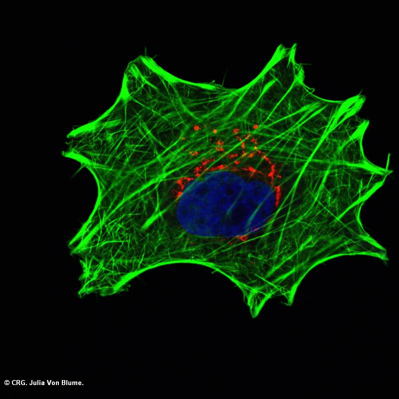
Title: HeLa cells
Ref: VM-0006
Author: JULIA VON BLUME
Size: 800 x 800
Description: Actin_TGN HeLa cells were transfected with ADF/cofilin siRNA for 48 h fixed and stained with phalloidin (green), TGN46 and Dapi (blue).
Explore CRG Images
The aim of the CRG photo gallery is to provide an overview of our scientists’ research activities through the images generated in the course of their research projects.
All our images are available on demand in digital form with higher quality for for-profit and any other uses. For any of these requests please contact us.
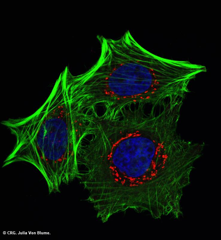
Title: HeLa cells
Ref: VM-0005
Author: JULIA VON BLUME
Size: 738 x 800
Description: Actin_TGN HeLa cells were transfected with ADF/cofilin siRNA for 48 h fixed and stained with phalloidin (green), TGN46 and Dapi (blue).
Explore CRG Images
The aim of the CRG photo gallery is to provide an overview of our scientists’ research activities through the images generated in the course of their research projects.
All our images are available on demand in digital form with higher quality for for-profit and any other uses. For any of these requests please contact us.
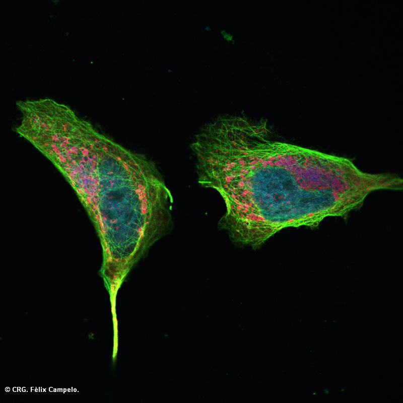
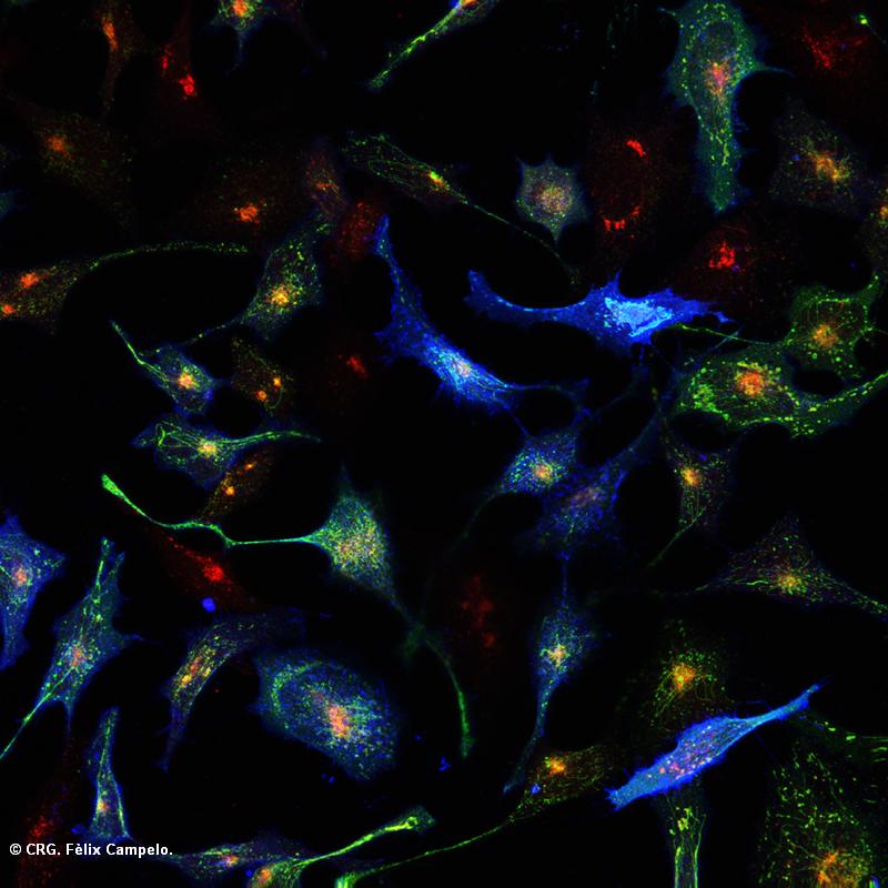
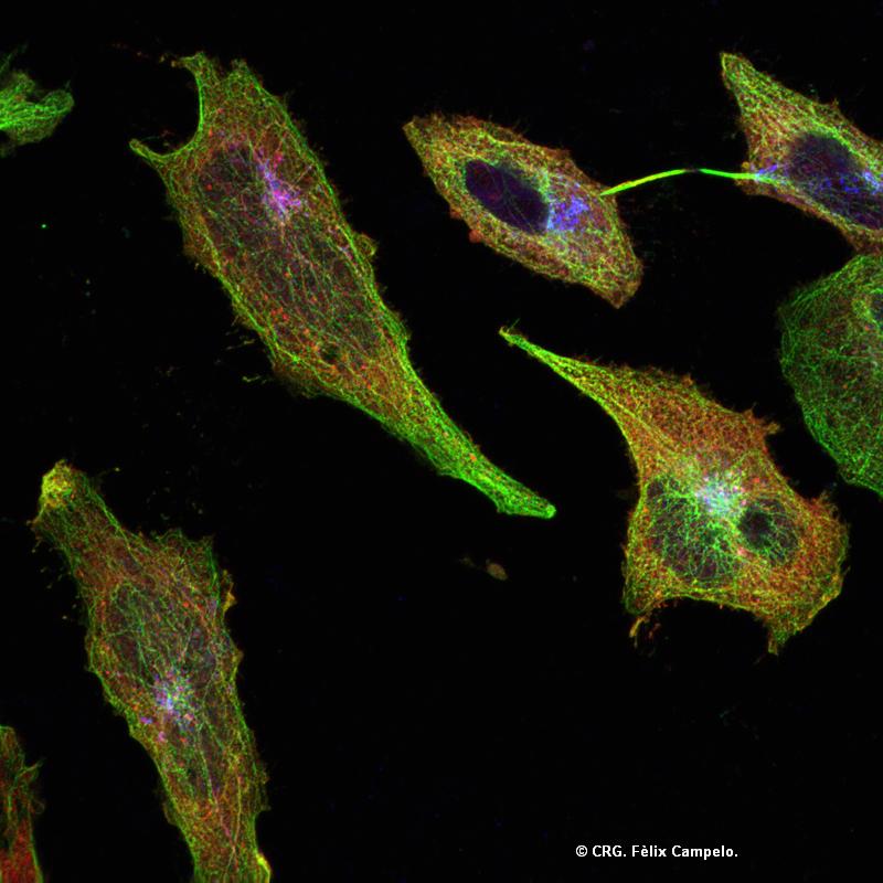
Title: HeLa cell
Ref: VM-0004
Author: FELIX CAMPELO
Size: 800 x 800
Description: HeLa cell showing the microtubule network and transport related proteins emanating from Golgi apparatus.
Explore CRG Images
The aim of the CRG photo gallery is to provide an overview of our scientists’ research activities through the images generated in the course of their research projects.
All our images are available on demand in digital form with higher quality for for-profit and any other uses. For any of these requests please contact us.
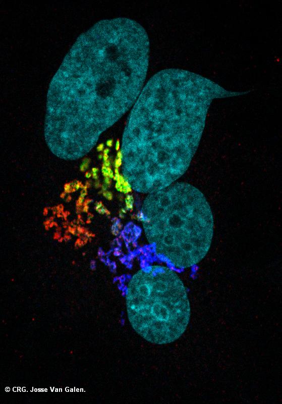
Title: Fused human and rat cells
Ref: VM-0003
Author: JOSSE VAN GALEN
Size: 559 x 800
Description: Fused rat and human cells stained for several Golgi markers: Red - GRASP65 (human) Green - Mannosidase-II-GFP (human) Blue - Mannosidase-II (rat) Cyan - Nucleus DNA
Explore CRG Images
The aim of the CRG photo gallery is to provide an overview of our scientists’ research activities through the images generated in the course of their research projects.
All our images are available on demand in digital form with higher quality for for-profit and any other uses. For any of these requests please contact us.
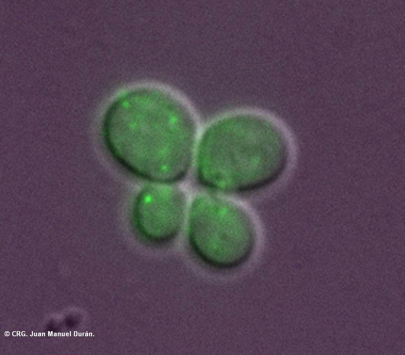
Title: Budding yeasts
Ref: VM-0002
Author: JUAN DURAN
Size: 800 x 700
Description: Two budding yeasts (S. cerevisiae) undergoing cell division next to each other. They are stained with a Golgi marker in green
Explore CRG Images
The aim of the CRG photo gallery is to provide an overview of our scientists’ research activities through the images generated in the course of their research projects.
All our images are available on demand in digital form with higher quality for for-profit and any other uses. For any of these requests please contact us.
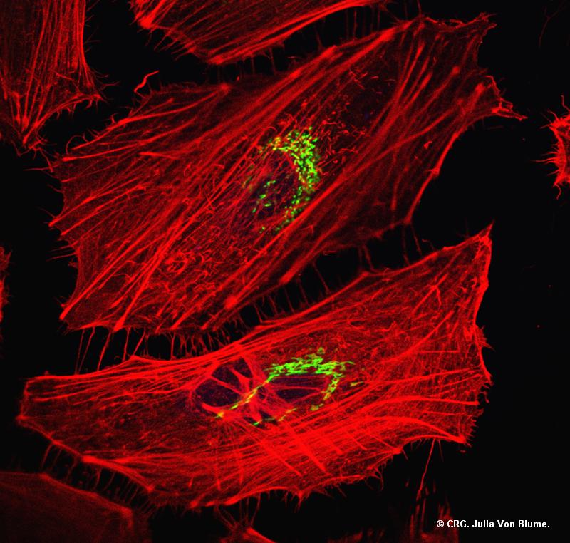
Title: Golgi apparatus in HeLa cells
Ref: VM-0001
Author: JULIA VON BLUME
Size: 800 x 762
Description: In green the Golgi apparatus and in red the actin cytoskeleton (HeLa cells)
Explore CRG Images
The aim of the CRG photo gallery is to provide an overview of our scientists’ research activities through the images generated in the course of their research projects.
All our images are available on demand in digital form with higher quality for for-profit and any other uses. For any of these requests please contact us.
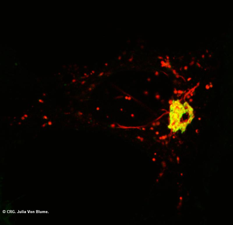
Title: Golgi apparatus transport carriers
Ref: VM-0012
Author: JULIA VON BLUME
Size: 800 x 773
Description: Formation of Transport carriers from the Golgi HeLa cells stably expressing Mann-2 GFP were transfected with ssRFP (soluble secretory protein), cargo was arrested in the TGN at 20°C and released at 32°C. The red dots and tubes represent TGN derived transport carriers.
Explore CRG Images
The aim of the CRG photo gallery is to provide an overview of our scientists’ research activities through the images generated in the course of their research projects.
All our images are available on demand in digital form with higher quality for for-profit and any other uses. For any of these requests please contact us.
Title: HeLa cells in culture
Ref: VM-0011
Author: FELIX CAMPELO
Size: 800 x 800
Description: Heterogeneous population of HeLa cells transfected with a protein kinase and one of its substrates showing different tubes emanating from the Golgi apparatus.
Explore CRG Images
The aim of the CRG photo gallery is to provide an overview of our scientists’ research activities through the images generated in the course of their research projects.
All our images are available on demand in digital form with higher quality for for-profit and any other uses. For any of these requests please contact us.
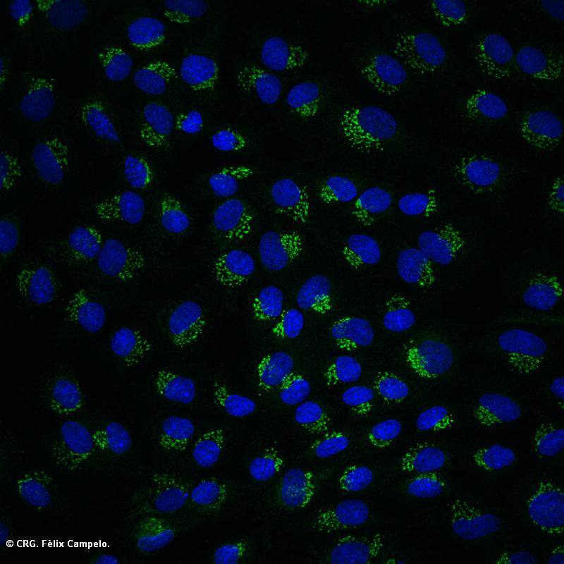
Title: Kidney cells
Ref: VM-0010
Author: FELIX CAMPELO
Size: 800 x 800
Description: Golgi apparatus (green) and nucleus (blue) of normal rat kidney cells.
Explore CRG Images
The aim of the CRG photo gallery is to provide an overview of our scientists’ research activities through the images generated in the course of their research projects.
All our images are available on demand in digital form with higher quality for for-profit and any other uses. For any of these requests please contact us.