Cellular & Systems Neurobiology
Explore CRG Images
The aim of the CRG photo gallery is to provide an overview of our scientists’ research activities through the images generated in the course of their research projects.
All our images are available on demand in digital form with higher quality for for-profit and any other uses. For any of these requests please contact us.
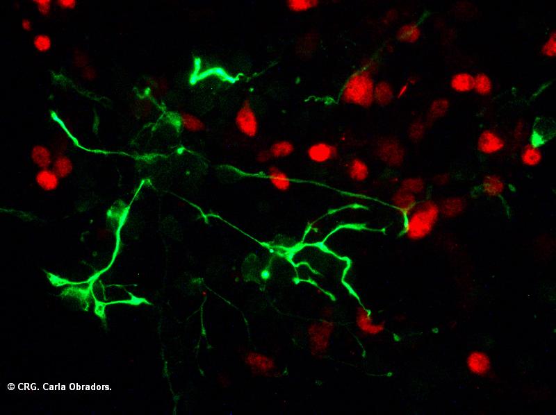
Title: Cortical neurons culture
Ref: MD-0002
Author: CARLA OBRADORS
Size: 800 x 598
Description: Mouse cortical neurons in culture. Neurons labeled in red (NeuN) and glial neurons in green (GFAP)
Explore CRG Images
The aim of the CRG photo gallery is to provide an overview of our scientists’ research activities through the images generated in the course of their research projects.
All our images are available on demand in digital form with higher quality for for-profit and any other uses. For any of these requests please contact us.
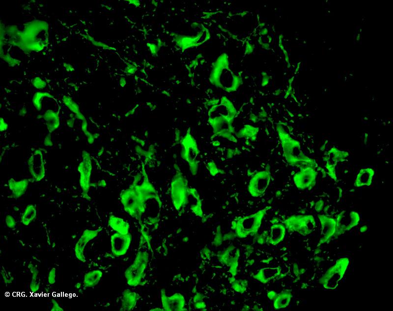
Title: Ventral tegmental area from mouse brain
Ref: MD-0004
Author: XAVIER GALLEGO
Size: 800 x 634
Description: Ventral tegmental area from mouse brain. Immunohistochemistry of tirosyn hidroxilase to label the dopaminergic neurons (green)
Explore CRG Images
The aim of the CRG photo gallery is to provide an overview of our scientists’ research activities through the images generated in the course of their research projects.
All our images are available on demand in digital form with higher quality for for-profit and any other uses. For any of these requests please contact us.

Title: Nucleus coeruleus from mouse brain
Ref: MD-0003
Author: XAVIER GALLEGO
Size: 800 x 634
Description: Nucleus Coeruleus from mouse brain. Immunihistochemistry of tirosyn hidroxilase to label the noradrenergic neurons (green)
Explore CRG Images
The aim of the CRG photo gallery is to provide an overview of our scientists’ research activities through the images generated in the course of their research projects.
All our images are available on demand in digital form with higher quality for for-profit and any other uses. For any of these requests please contact us.

Title: Nucleus coeruleus from mouse brain
Ref: MD-0001
Author: XAVIER GALLEGO
Size: 800 x 800
Description: Nucleus Coeruleus from mouse brain. Immunihistochemistry of TrKC (neurothophin receptor NT-3) (red) and tirosyn hidroxilase to label the noradrenergic neurons (green)
Mara Dierssen
Research and Interests
Dr. Dierssen is interested in brain mechanisms underlying learning and memory and how these are altered in pathological cognitive states. Her research focuses on understanding how neural architecture and connectivity constrain mesoscopic network activity and influences the flow and storage of information in neural circuits in human cognitive-related disorders. More specifically, current research lines encompass functional connectivity in intellectual disability (e.g. Down syndrome); mechanisms underlying reconsolidation of traumatic fear memories in panic disorder; working memory and impulsivity in relation to sensitivity to develop nicotine dependence; and a phenotyping roadmap of obesity pandemics.
Retrieving and consolidation of memory is a key capability of the human brain, necessary for normal daily functioning. Improved understanding of their mechanisms can provide a major contribution to new and improved therapies in cognitive disorders.