Explore CRG Images
The aim of the CRG photo gallery is to provide an overview of our scientists’ research activities through the images generated in the course of their research projects.
All our images are available on demand in digital form with higher quality for for-profit and any other uses. For any of these requests please contact us.

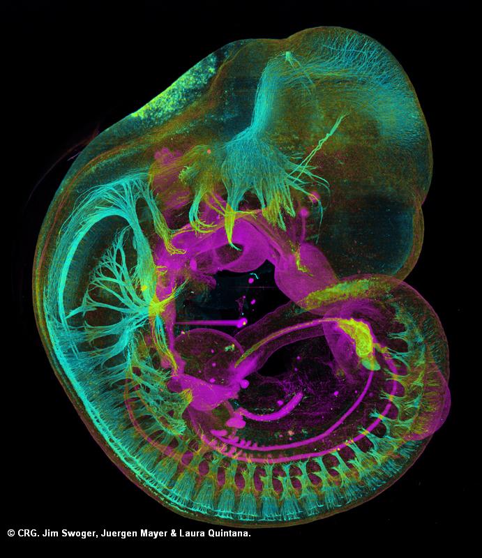
Title: Mouse Embryo with Neurofilament and E-Cadherin Staining
Ref: JS-0060
Author: Jim Swoger, Juergen Mayer, Laura Quintana
CRG Group: Multicellular Systems Biology
Size: 691 x 800
Description: Structures containing Neurofilament (cyan) and E-cadherin (magenta) are visualized in the embryo, highlighting structures in the developing nervous system and internal organs. The image is a maximum-value projection through the 3D data set generated by the microscope. To achieve high resolution, 12 sub-regions of the sample were scanned and combined into a montage.

